Plasma Cell Disorders (Dyscrasias)
Plasma Cell Disorders Pathology Video
Plasma cell disorders are also known as plasma cell dyscrasias.
Examples of plasma cell disorders include:
- Multiple myeloma (MM)
- Monoclonal gammopathy of undetermined significance (MGUS)
- Waldenstrom macroglobulinemia (WM)
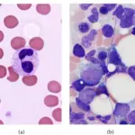
 Multiple Myeloma. (a and b) Histopathological appearance of bone marrow biopsy displaying plasmacytosis (H and E, ×40). Localized cutaneous mucinosis associated with multiple myeloma: a rare presentation. Rather PA, Hussain M, Bagdadi F - Indian journal of dermatology (2014). Not altered. CC.

Multiple Myeloma
Multiple myeloma (MM) is a malignant proliferation of plasma cells.
Multiple myeloma (MM) requires greater than 10% of the bone marrow cells to be malignant plasma cells.
Multiple myeloma (MM) is the most common primary malignancy of bone.
The neoplastic plasma cells produce immunoglobulin.
Multiple myeloma (MM) is associated with high serum levels of IL-6.
The excess immunoglobulin can be detected on serum protein electrophoresis (SPEP), resulting in a monoclonal spike (M spike), most commonly due to monoclonal IgG or IgM.
Osteoclasts that have the RANK receptor are activated by neoplastic plasma cells, and characteristically degrade the bone.
Clinical and laboratory findings of multiple myeloma (MM) include:
- Bone pain with hypercalcemia
- Lytic, ‘punched-out’ skeletal lesions are seen on x-ray, especially in the vertebrae and skull
- Fractures after minor trauma
- Elevated serum protein
- Increased risk of infections
- Rouleaux formation of red blood cells (RBCs) on blood smear due to increased serum protein decreasing charge between RBCs
- Primary AL amyloidosis in which kappa free light chains circulate in serum and deposit in tissues
- Proteinuria due to kappa or lambda free light chains being excreted in the urine as Bence Jones protein
Monoclonal antibodies of multiple myeloma lack antigenic diversity.
The most common cause of death in patients with multiple myeloma is infection.
Bence Jones protein deposits in kidney tubules, increasing the risk of kidney failure.
Kidney failure secondary to multiple myeloma is myeloma kidney.
 Multiple Myeloma. Aspiration cytology smear of a case plasmacytoma in 4 cm diameter chest wall mass in a 69 year male patient. (MGG stain, ×440). Cytology of plasma cell rich effusion in cases of plasma cell neoplasm. Journal of Cytology / Indian Academy of Cytologists. Not altered. CC.


Monoclonal Gammopathy of Undetermined Significance (MGUS)
Monoclonal gammopathy of undetermined significance (MGUS) is an increased serum protein with M spike on SPEP.
MGUS is common in the elderly population.
Approximately 5% of individuals above 70-years-old have monoclonal gammopathy of undetermined significance (MGUS).
One percent of patients with monoclonal gammopathy of undetermined significance (MGUS) develop multiple myeloma each year.
 MGUS. a: Densitometry tracing of gel electrophoresis of normal serum.b: Densitometric tracing of gel electrophoresis of subject with monoclonal band. Prevalence and type of monoclonal gammopathy of undetermined significance in an apparently healthy Nigerian population: a cross sectional study. Onwah AL, Adeyemo TA, Adediran A, Ajibola SO, Akanmu AS - BMC blood disorders (2012). Not altered. CC.


Waldenstrom Macroglobulinemia
Waldenstrom macroglobulinemia (WM) is B-cell lymphoma with monoclonal IgM production.
Clinical features of Waldenstrom macroglobulinemia (WM) include:
- Lymphadenopathy
- Increased serum protein with M spike (comprised of lgM)
- Visual deficits
- Symptoms related to hyperviscous blood due to the IgM (large pentamer) causing serum hyperviscosity
- Neurologic deficits such as hemorrhage or stroke
- Bleeding due to viscous serum results in defective platelet aggregation
Acute complications are treated with plasmapheresis, which removes lgM from the serum.
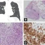
 Waldenstrom Macroglobulinemia. The tissue or bone marrow involvement pattern and immunophenotypic profiling of the EMWM and BMWM patients. a, b A representative diffuse interstitial pattern of bone marrow biopsy in a patient with BMWM, ×40 and × 80 magnification. c Bone marrow aspirate smear showed many abnormal small lymphoid cells admixed with variable plasmacytoid cells and plasma cells, ×80 magnification. d CD20 stain on the abnormal small lymphoid cells, ×80 magnification. e CD138 stain on the plasmacytoid and plasma cells, ×80 magnification. f, g Immunophenotypic profiling of CD38 and CD138 positive plasmacytoid and plasma cells with strong monotypic cytoplasmic kappa light chain expression. h The abnormal small lymphoid cells were CD20 positive and showed monotypic kappa light chain expression. i, j A representative diffuse infiltrative pattern of a lymph node biopsy in a patient with EMWM, MYD88 mutation positive, ×40 and × 80 magnification. k CD20 stain on abnormal small lymphoid cells, ×80 magnification. l CD138 stain on few admixed plasmacytoid and plasma cells, ×80 magnification. m, o Immunophenotypic profiling of CD19 and CD20 positive small B-cells with kappa light chain restriction and were negative for CD5 and CD10. p Identical monotypic kappa light chain expression in the lymphoid cells was also seen in the plasmacytoid and plasma cells. Waldenström macroglobulinemia with extramedullary involvement at initial diagnosis portends a poorer prognosis. Cao X, Ye Q, Orlowski RZ, Wang X, Loghavi S, Tu M, Thomas SK, Shan J, Li S, Qazilbash M, Yin CC, Weber D, Miranda RN, Xu-Monette ZY, Medeiros LJ, Young KH - Journal of hematology & oncology (2015). Not altered. No license or rights.