Arteriosclerosis, Aortic Dissection, and Aneurysm Pathology Video
Arteriosclerosis
Arteriosclerosis is due to the blood artery wall thickening, literally “hard arteries.”
The types of arteriosclerosis include:
- Atherosclerosis (hardening and narrowing of large arteries)
- Arteriolosclerosis (hardening and calcification of small arteries)
- Mönckeberg medial calcific sclerosis (hardening and narrowing of small to medium sized arteries)
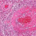
 Arteriolosclerosis. "Onion-skin" renal arteriole Pacolarosa - Not altered. CC BY-SA 3.0


Atherosclerosis
Atherosclerosis causes blood flow is obstructed by intimal plaque.
Atherosclerosis typically involves medium-sized and big arteries.
Atherosclerosis frequently develops dystrophic calcification.
Arteries commonly affected by Atherosclerosis include:
- Abdominal aorta
- Internal carotid artery
- Popliteal artery
There are two categories of risk factors for atherosclerosis:
- Modifiable
- Non-modifiable
Modifiable risk factors of atherosclerosis include:
- Hypertension
- Hypercholesterolemia (LDL raises risk, HDL lowers risk)
- Smoking
- Diabetes
Non-modifiable risk factors of atherosclerosis include:
- Age
- Gender (men > women)
- Genetics
Men and postmenopausal women are at increased risk.
Estrogen is protective against atherosclerosis, thus menopause is a risk factor as well.
The frequency and severity of atherosclerosis increases with age.
Pathogenesis of atherosclerosis involves:
- Lipids leak into the tunica intima as a result of endothelial damage
- Foam cells are created when oxidized lipids are taken up by macrophages through scavenger receptors
- Extracellular matrix is deposited during inflammation and healing, which also promotes the growth of smooth muscle
Histologically, atherosclerosis consists of a fibromuscular cap covering a necrotic lipid core (mainly cholesterol).
Morphologic stages of atherosclerosis include:
- Fatty streaks
- Plaques
Atherosclerosis is initialized as fatty streaks (flat yellow lesions of the intima consisting of lipid-laden macrophages).
The fatty streaks tend to presents early in life for the majority of teenagers.
The development of atherosclerotic plaque ensues over young adulthood to elder years.
Over 50% of diseases in Western nations are complications of atherosclerosis.
Complications of atherosclerosis include:
- Peripheral vascular, which disease develops as a result of ischemia and reduced blood flow caused by medium-sized artery stenosis (lower extremity arteries, e.g., popliteal)
- Angina (coronary arteries)
- Irritable bowel syndrome (mesenteric arteries)
- Myocardial infarction (coronary arteries) and stroke are caused by plaque rupture with thrombosis (e.g., middle cerebral artery)
- Atherosclerotic emboli, which are identified by cholesterol crystals within the embolus, are the result of plaque rupture and embolization
- Aneurysms are caused by the vessel wall weakening (e.g., abdominal aorta)
 Arteriosclerosis. Gross pathology of atherosclerosis, aorta. Autopsy specimen of aorta has been opened lengthwise to reveal luminal surface studded with lesions of atherosclerosis. Content Providers(s): CDC/Dr. Edwin P. Ewing, Jr. Creation Date: 1972. Not altered. CC.
Arteriolosclerosis
Arteriolosclerosis results in small arteriole narrowing.
Arteriolosclerosis is categorized as hyaline and hyperplastic varieties.
Protein leakage into the arterial wall, which results in vascular thickening, is the primary cause of hyaline arteriolosclerosis.
On microscopy arteriosclerosis appears as pink hyaline around the circumference of the vessel.
Risk factors for arteriolosclerosis include:
- Hypertension
- Diabetes
Arteriolosclerosis may results in end-organ ischemia and reduced vascular diameter such as causes glomerular scarring (arteriolonephrosclerosis), which advances gradually to chronic renal failure.
Hyperplastic arteriolosclerosis thickens the artery wall by causing smooth muscle hyperplasia in an “onion-skin” pattern.
Fibrinoid necrosis of the vessel wall may also be seen in hyperplastic arteriolosclerosis.
Malignant hypertension may be related to hyperplastic arteriosclerosis due to rapid decreased vessel diameter.
Arteriolosclerosis may cause acute renal failure, and has a distinctive “flea-bitten” appearance.
- Arteriolosclerosis. "Onion-skin" renal arteriole Pacolarosa - Not altered. CC BY-SA 3.0
Mönckeberg Medial Calcific Sclerosis
Mönckeberg medial calcific sclerosis results in the calcification of the muscular tunica media portion of medium-sized arteries.
Mönckeberg medial calcific sclerosis is non-obstructive.
Mönckeberg medial calcific sclerosis is typically not clinically significantly.
Mönckeberg medial calcific sclerosis is usually an unintended discovery in X-ray mammography.
 Monckeberg Medial Calcific Sclerosis. A. Pelvic and lower extremity radiograph shows extensive calcification of the femoral arteries. B. Translumbar aortography shows near-total obstruction of the femoral arteries. Couri CE, da Silva GA, Martinez JA., Pereira Fde A, de Paula FJ. - Couri CE, da Silva GA, Martinez JA., Pereira Fde A, de Paula FJ. Mönckeberg's sclerosis Not altered. CC BY 2.0

Aortic Dissection
Aortic dissection is caused by shearing force allowing blood leaking through the media of the aortic wall due to an intimal tear.
Aortic dissection occurs in the high-stress zone of the proximal portion of the aorta and is accompanied by medial weakening.
Risk factors of aortic dissection include:
- Hypertension in elderly persons, characterized by vasa vasorum hyaline arteriosclerosis
- Genetic connective tissue abnormalities in younger individuals such as Marfan syndrome or Ehlers-Danlos syndrome, characterized by weak connective tissue and cystic medial degeneration
The media sags as a result of decreased flow.
Weakness of the connective tissue in the media is a characteristic symptom of Marfan syndrome and Ehlers-Danlos syndrome (cystic medial necrosis).
Symptoms of aortic dissection include:
- Intense chest pain
- Chest pain that radiates to the back
Complications of aortic dissection include:
- Death secondary to cardiac tamponade (the most common)
- Rupture with lethal hemorrhage
- Obstruction of branching arteries (such as the coronary or renal arteries)
- End-organ ischemia
 Aortic Dissection. (A) Computed tomography image showing retrograde type A aortic dissection (arrow) at three months after endovascular repair and (B) last follow-up image at twenty months after the ascending aorta replacement. Surgical repair of retrograde type a aortic dissection after thoracic endovascular aortic repair. Kim CY, Chang WI, Kim YS, Ryoo JY - The Korean journal of thoracic and cardiovascular surgery (2014). Not altered. CC.


Aneurysm
Thoracic Aneurysm
An aneurism of the thoracic aorta is a thoracic aneurysm.
Thoracic aneurysm risk factors include:
- Syphilis
- Hypertension
Tertiary syphilis is a common cause of thoracic aneurysm.
Tertiary syphilis can cause vasa vasorum endarteritis leading to thoracic aneurysm due to:
- Luminal constriction
- Reduced blood flow
- Vascular wall atrophy
Tertiary syphilis results in the aorta having a “tree-bark” look.
Complications of thoracic aneurysm includes:
- Airway compression
- Esophagus compression
- Thrombosis
- Embolism
 Thoracic Aneurysm. A stent graft placed in the thoracic aorta to treat a thoracic aortic aneurysm. James Heilman, MD - Not altered. CC BY-SA 3.0


Abdominal Aortic Aneurysm
Abdominal aortic aneurysm (AAA) is an aneurysm of the abdominal aorta.
Abdominal aortic aneurysm typically develops above the aortic bifurcation but below the renal arteries.
Abdominal aortic aneurysm (AAA) is typically associated with hypertension and atherosclerosis.
Risk factors for abdominal aortic aneurysm (AAA) include:
- Male sex
- Smoking
- Elderly
- Atherosclerosis
- Hypertension
Atherosclerosis causes the vessel wall to atrophy and become weaker by raising the diffusion barrier to the media.
Abdominal aortic aneurysm (AAA) typically presents with a triad of:
- Flank pain
- Pulsating abdominal mass
- Hypotension
Symptoms of abdominal aortic aneurysm include an abdomen lump that pulses initially and enlarges over time.
Complications of abdominal aortic aneurysm (AAA) include:
- Rupture, especially when the diameter is greater than 5 cm
- Compression of nearby structures (such as the ureter)
- Thrombosis
- Embolism
 Abdominal Aortic Aneurysm. Abdominal aortic aneurysms (3.4 cm) Intermedichbo at Serbian Wikipedia Abdominal aortic aneurysm 34 мм, Author: Milorad Dimic MD, Nis, Srbia, 2009. Not altered. CC BY-SA 3.0 rs