Small bowel pathology includes:
- Duodenal atresia
- Meckel diverticulum
- Volvulus
- Intussusception
- Small bowel infarction
- Lactose intolerance
- Celiac disease
- Tropical sprue
- Whipple disease
- Abetalipoproteinemia
- Neuroendocrine tumor
- Acute appendicitis
Duodenal Atresia
Duodenal atresia is due to a congenital failure of duodenum to canalize.
Imaging will classically show a double bubble sign in duodenal atresia.
Duodenal atresia is linked to Down syndrome.
Clinical features of duodenal atresia include:
- Polyhydramnios
- Stomach distension and a duodenal loop that is blind (the “double bubble” indication)
- Bilious vomiting
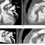
 Duodenal Atresia. Duodenal Atresia Neonate Kinderradiologie Olgahospital Klinikum Stuttgart. Not altered. CC BY-SA 3.0
Meckel Diverticulum
Meckel diverticulum is when the gut wall’s three layers are all outpouched (true diverticulum).
Meckel diverticulum occurs because of the failure of the vitelline duct to involute.
Meckel diverticulum is associated with the rule of twos:
- 2% of the population
- 2% are symptomatic
- 2 years old or younger
- 2:1 male to female ratio
- 2 feet proximal to the ileocecal valve
- 2 inches long
- 2 types of mucosal lining
 Meckel Diverticulum. Meckel's diverticulum surgical specimen Unknown author - Armed Forces Institute of Pathology Not altered. Public Domain


Volvulus
Volvulus is twisting of bowel along its mesentery.
Volvulus can lead to obstruction and disruption of the blood supply with infarction (mesenteric ischemia).
Common locations of volvulus are:
- Sigmoid colon (elderly)
- Cecum (young adults)
 Volvulus. Plain X ray of a cecal volvulus James Heilman, MD - Not altered. CC BY-SA 4.0


Intussusception
Intussusception is telescoping of the proximal portion of the bowel into the distal portion.
Peristalsis pulls the telescoped segment forward, which causes blockage and interruption of the blood supply with infarction.
In children, lymphoid hyperplasia is the most common cause of intussusception.
Intussusception typically occurs in the terminal ileum, resulting in intussusception into the cecum.
The most prevalent cause of intussusception in adults is a tumor.
 Intussusception. Drawing of intussusception Olek Remesz (wiki-pl: Orem, commons: Orem) - Not altered. CC BY-SA 3.0


Small Bowel Infarction
Small bowel damage from ischemia can be very serious.
Transmural infarction presents with thrombosis/embolism of the superior mesenteric artery or thrombosis of the mesenteric vein.
Mucosal infarction presents with marked hypotension.
Features of small bowel infarction include:
- Abdominal pain
- Bloody diarrhea
- Decreased bowel sounds
 Small Bowel Infarction. Bowel infarction. Note the grey discoloration. haitham alfalah. Not altered. CC BY-SA 3.0

Lactose Intolerance
Reduction in the lactase enzyme’s activity, which is located at the brush boundary of enterocytes.
Lactase typically breaks down lactose into glucose and galactose.
By consuming milk products undigested lactose is osmotically active and causes stomach distension and diarrhea.
Lactose deficiency can be congenital (rare autosomal recessive disorder) or acquired (usually occurs in late childhood).
Temporary deficiency of lactose is visible after small bowel infection (lactase is highly susceptible to injury).
 Lactose Intolerance. Skin prick testing for allergies. For a positive response, the skin will become red and raised. National Institutes of Health (NIH) - National Institutes of Health (NIH). Not altered. Public Domain
Celiac Disease
Celiac disease is an immune-mediated damage of small bowel villi because of gluten exposure.
Celiac disease is linked to HLA-DQ2 and HLA-DQ8.
Wheat and other cereals contain the protein gluten, whose pathogenic component is gliadin.
Tissue transglutaminase deamidates gliadin after it has been absorbed (TTG).
Antigen-presenting cells use MHC class I to present deaminated gliadin.
Tissue injury is mediated by helper T lymphocytes.
Clinical presentation of celiac disease includes:
- Bloating
- Diarrhea
- Undernutrition
- Small, herpes-like vesicles can be visible on skin called dermatitis herpetiformis
Symptoms of celiac disease resolve with a gluten-free diet.
Laboratory findings of celiac disease include:
- IgA antibodies against endomysium
- Tissue transglutaminase (tTG)
- Gliadin
They are also present and are helpful for diagnosing people with IgA deficiency (higher incidence of IgA deficiency is visible in celiac disease).
Histology of celiac disease shows:
- IgA deposition at the tips of dermal papillae
- Villi are flattened, crypts are hyperplastic, and there are more intraepithelial lymphocytes, according to a duodenal biopsy
The jejunum and ileum are less affected than the duodenum, which has the most obvious damage.
Complications of celiac disease include:
- Small bowel carcinoma
- T-cell lymphoma
 Celiac Disease. Immunofluorescence staining pattern of endomysial antibodies on a monkey oesophagus tissue sample. Simon Caulton - Not altered. CC BY-SA 3.0


Tropical Sprue
Tropical sprue is damage to small bowel villi because of an unknown organism, leading to malabsorption.
Comparable to celiac disease, but can only be seen in tropical areas such as the Caribbean.
Tropical sprue occurs after infectious diarrhea and is treated with antibiotics.
The jejunum and ileum are most typically affected, and secondary vitamin B12 or folate insufficiency may result.
The duodenum is less frequently affected by tropical sprue.
 Duodenal biopsy of a patient with tropical sprue. Partial villous atrophy (arrow) and lymphoplasmacytic infiltration in the lamina propria (asterisk) are visible. The crypt to villous ratio is almost 1:1 (H&E, ×20). Spectrum of chronic small bowel diarrhea with malabsorption in Indian subcontinent: is the trend really changing? Pipaliya N, Ingle M, Rathi C, Poddar P, Pandav N, Sawant P - Intestinal research (2016). Not Altered. CC.
Whipple Disease
Whipple disease is a systemic tissue injury characterized by Tropheryma Whipplei (formerly called Tropheryma Whippelii) organisms.
Organisms that have partially been killed are found in macrophage lysosomes (positive for PAS).
The small bowel lamina propria is the classic site of involvement.
Macrophages compress lacteals.
Chylomicrons can’t be moved from enterocytes to lymphatics.
Whipple disease leads to fat malabsorption and steatorrhea.
Other usual sites of involvement of Whipple disease include:
- Synovium of joints causing arthritis
- Cardiac valves
- Central nervous system
- Lymph nodes
 Whipple Disease. High magnification micrograph showing the characteristic foamy macrophages in the lamina propria, H&E stain Nephron -Not altered. CC BY-SA 3.0

Abetalipoproteinemia
Apolipoproteins B-48 and B-100 deficiency are autosomal recessive conditions that can cause abetalipoproteinemia.
Clinical features of abetalipoproteinemia include:
- Malabsorption because of defective chylomicron formation (needs B-48)
- Absent plasma VLDL and LDL (need B-100)
 Abetalipoproteinemia. Very high magnification micrograph of abetalipoproteinemia. Duodenal biopsy. H&E stain. The small bowel mucosa shows the characteristic clear enterocytes (due to lipid accumulation). Brunner's glands are not seen on the images. Related images Intermed. mag. High mag. Very high mag. Nephron. Not altered. CC BY-SA 3.0


Neuroendocrine Tumor
Neuroendocrine tumor is also known as carcinoid tumor.
Neuroendocrine tumors are malignant proliferations of neuroendocrine cells.
Neuroendocrine tumors are positive for CD56, synaptophysin, chromogranin.
Neuroendocrine tumors can occur anywhere in the gut, with the small bowel being the most typical place.
Neuroendocrine tumors typically begin as small nodules that resembles a submucosal polyp.
Neuroendocrine tumors frequently secrete serotonin.
Serotonin is released into the bloodstream through the portal vein and converted to 5-HIAA by the liver monoamine oxidase (MAO).
The urine contains excretion of 5-HIAA.
Serotonin can bypass liver metabolism when a carcinoid tumor spreads to the liver.
Carcinoid syndrome and carcinoid heart disease are caused by serotonin being released into the hepatic vein and leaking into the systemic circulation via hepato-systemic shunts.
Symptoms of carcinoid syndrome include:
- Bronchitis
- Diarrhea
- Skin flushing
These symptoms of carcinoid syndrome can be brought on by:
- Eating cured meats
- Alcohol consumption (i.e. red wine)
- Mental stress
Due to the presence of monoamine oxidase, which metabolizes serotonin in the lung, left-sided valvular lesions are not visible.
Carcinoid syndrome is characterized by right-sided valvular fibrosis (increased collagen) that causes tricuspid regurgitation and pulmonary valve stenosis.
") Well-differentiated neuroendocrine tumor of the duodenum. Ed Uthman. Not altered. CC BY 2.0
Well-differentiated neuroendocrine tumor of the duodenum. Ed Uthman. Not altered. CC BY 2.0
")

Acute Appendicitis
Acute appendicitis is due to acute inflammation of the appendix.
Acute appendicitis is the most prevalent cause of acute abdomen.
Acute appendicitis is commonly associated with a fecalith or lymphoid hyperplasia that is obstructing the appendix.
Clinical features of acute appendicitis include:
- Periumbilical pain
- Fever
- Nausea
- Pain subsequently localizes to the right lower quadrant (McBurney point)
- Guarding and rebound tenderness are symptoms of peritonitis, which occurs as a result of rupture
Complications of acute appendicitis like periappendiceal abscess are frequent.
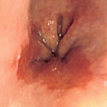
 Acute Appendicitis. An exemplary case of acute appendicitis in a 10-year-old boy. The organ is enlarged and sausage-like (botuliform). This longitudinal section shows the angry red inflamed mucosa with its irregular luminal surface. Diagnosed and removed early in the course of the disease, this appendix does not show late complications, like transmural necrosis, perforation, and abscess formation. This photo was shot with a Nikon Coolpix 8800 with no macro attachment or other accessory. I used the "Closeup" setting in the "Scene" mode after presetting the white balance against a standard grey card. The specimen is slightly raised over a black velvet background and illuminated by Photofloods. Editing, done with Picasa, consisted of cropping, sharpening, and adjusting levels. This is an example of one of my "quick 'n' dirty" specimen photos, requiring little time and equipment but still yielding acceptable results. If I were to shoot the same specimen for publication or competition, I would use a digital SLR with dedicated macro lens, spend more time preparing and propping up the specimen (to maximize the area in focus), take at least a dozen shots (rather than the two done for this one) with exposure bracketing, and do all editing in Photoship on the Mac at home (where I don't get a zillion interruptions). 😉 Ed Uthman. Not altered. CC BY 2.0