Urinary Tract Infections Pathology Video
Urinary Tract Infections
Urinary tract infections are infection of the urethra, bladder, or kidney.
Urinary tract infections include:
- Cystitis
- Sterile pyuria
- Urethritis
- Pyelonephritis
- Chronic pyelonephritis
- Nephrolithiasis
Ascending infection is the primary cause of urinary tract infections (UTIs), and females are more likely to develop it than males.
Risk factors for urinary tract infections (UTIs) include:
- Urinary stasis
- Multiple sexual partners
- Urinary catheter use
- Congenital abnormalities of the urinary tract
 Urinary Tract Infections. Uropathogenic Escherichia coli (UPEC) cells adhered to bladder epithelial cell. Stefan Walkowski - Not altered. CC BY-SA 4.0


Cystitis
Cystitis is bladder infection.
Symptoms of cystitis include:
- Fever
- Dysuria
- Urinary frequency
- Urinary urgency
- Suprapubic pain
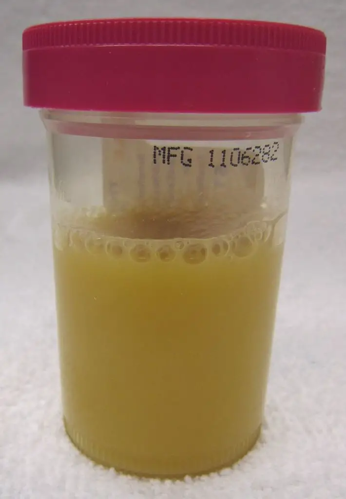
Laboratory results of cystitis include:
- Urinalysis-cloudy urine with greater than 10 white blood cells (WBCs) per high power field (HPF)
- Dipstick-positive leukocyte esterase (because of pyuria) and nitrites (bacteria converts nitrates into nitrites)
- Culture-greater than 100,000 colony-forming units (gold standard)
Etiologies of cystitis include:
- E. coli (80% of cases)
- Staphylococcus saprophyticus in young, sexually active women (but E. Coli is still very usual in this population)
- Klebsiella pneumoniae
- Proteus mirabilis
- Enterococcus faecalis
 Chronic Cystitis. Chronic bladder infection in C3H/HeJ mice is histologically distinct from chronic cystitis in C3H/HeOuJ mice.Representative paraffin-embedded, fixed bladder sections from C3H/HeOuJ mice with chronic cystitis (22 out of 24 mice; panels A, D, & G), C3H/HeJ mice with high bacterial burdens and evidence of bladder inflammation (5 out of 20 mice; panels B, E, & H), and the remaining C3H/HeJ mice that uniformly lacked evidence of bladder inflammation (15 out of 20 mice; panels C, F, & I) were analyzed at 4 wpi; Bars approximate 50 µm in length. A–F, Sections were stained with hematoxylin & eosin and examined by light microscopy; boxes in panels A–C demarcate boundaries of higher magnification images in panels D–F. G–I, Sections were analyzed by indirect immunofluorescence (IF) microscopy, staining for E. coli (green) and uroplakin III (red), with nuclei counterstained with bis-benzimide (blue). “L” denotes bladder lumen; dashed line indicates approximate location of basement membrane; arrowheads point to urothelial surface bacterial colonization in panel E; and the white arrow points to a mature superficial facet cell containing a possible IBC in panel H. Early severe inflammatory responses to uropathogenic E. coli predispose to chronic and recurrent urinary tract infection. Hannan TJ, Mysorekar IU, Hung CS, Isaacson-Schmid ML, Hultgren SJ - PLoS pathogens (2010). Not Altered. CC.


Sterile Pyuria
Pyuria with a negative urine culture and more than 10 WBCs/HPF and leukocyte esterase is referred to as sterile pyuria.
 Sterile Pyuria. Pyuria in a person with urosepsis James Heilman, MD - Not altered. CC BY-SA 3.0

Urethritis
Urethritis is typically caused by Neisseria gonorrhoeae or Chlamydia trachomatis.
Dysuria is a common urethritis presenting symptom.
 Computed tomography of abdomen and pelvis showing (a) extensive edema and stranding of subcutaneous tissue at the symphysis pubis and bilateral groin areas; bilateral enlargement of inguinal lymph nodes is also noted (two arrows). (b) Edema and fat stranding extending into subcutaneous tissue at anterior lower abdominal wall area (two arrows).Fournier's Gangrene in a Heterosexual Man: A Complication of Neisseria meningitidis Urethritis. Khemees TA, Porshinsky BS, Patel AP, McClung CD - Case reports in urology (2012). Not Altered. CC.
Pyelonephritis
Pyelonephritis is kidney infection.
Pyelonephritis is usually brought on by ascending infection.
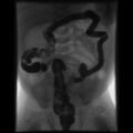
Vesicoureteral reflux is a risk factor of pyelonephritis.
Pyelonephritis presents with:
- Leukocytosis
- Fever
- Flank discomfort
- White blood cell casts
The most common pathogens that cause pyelonephritis include:
- E. Coli (90% of cases)
- Klebsiella species
- Enterococcus faecalis
Tubule atrophy and interstitial fibrosis as a result of repeated bouts of acute pyelonephritis.
Vesicoureteral reflux is the common cause of pyelonephritis in children.
Obstruction is a common cause of pyelonephritis in adults.
 Pyelonephritis. Acute pyelonephritis with increased cortical echogenicity and blurred delineation of the upper pole. Kristoffer Lindskov Hansen, Michael Bachmann Nielsen and Caroline Ewertsen -Not altered. CC BY 4.0
Chronic Pyelonephritis
Chronic pyelonephritis results in cortical scarring with blunted calyces.
Urinalysis may show waxy casts if a patient has chronic pyelonephritis.
Histology of chronic pyelonephritis may show atrophic tubules with eosinophilic proteinaceous debris that resembles thyroid follicles termed “thyroidization” of the kidney.
 Pyelonephritis. Chronic pyelonephritis with reduced kidney size and focal cortical thinning. Measurement of kidney length on the US image is illustrated by ‘+’ and a dashed line.[20] Kristoffer Lindskov Hansen, Michael Bachmann Nielsen and Caroline Ewertsen - (2015). Not altered.CC BY 4.0
Nephrolithiasis
Nephrolithiasis is the formation of a stone from a urine solute.
Risk factors for nephrolithiasis include:
- Low urine volume
- High solute concentration in the urinary filtrate
Nephrolithiasis presents as:
- Colicky pain
- Hematuria
- Tenderness on one flank
Stone(s) normally passes within a few hours.
If the stone does not pass by itself, medical or surgical intervention may be necessary.
 Nephrolithiasis. Renal ultrasonograph of a stone located at the pyeloureteric junction with accompanying hydronephrosis. Kristoffer Lindskov Hansen, Michael Bachmann Nielsen and Caroline Ewertsen - Not altered. CC BY 4.0