Pulmonary Infections Pathology Video
Pulmonary Infections
Pulmonary infections are disease of the lungs due to microorganisms.
Pulmonary infections include:
- Pneumonia (various types)
- Tuberculosis
Pneumonia
Pneumonia is a parenchymal lung infection.
Pneumonia may occur when the body’s natural defenses are weakened (e.g. impaired cough reflex, damage to mucociliary escalator, or mucus plugging).
Symptoms of pneumonia include:
- Fever
- Chills
- Productive cough that with yellow-green (pus) or rusty colored (bloody) sputum
- Tachypnea
- Pleuritic chest discomfort
Medical and lab findings of pneumonia include:
- Diminished breath sounds
- Dullness to percussion
- Elevated WBC count
- Chest X-ray
- Sputum gram stain and culture
Blood cultures are used to make the diagnosis of pneumonia.
Three common patterns of pneumonia may be detected on chest X-ray which include:
- Lobar pneumonia
- Bronchopneumonia
- Interstitial pneumonia
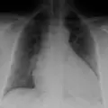
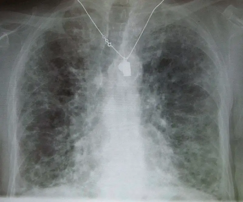
 Pneumonia. A chest x-ray of a patient with severe viral pneumonia due to SARS Not altered. Public Domain


Lobar Pneumonia
Lobar pneumonia is characterized by the consolidation of a whole lung lobe.
Lobar pneumonia is typically caused by bacteria which include:
- Streptococcus pneumonia (95 percent)
- Klebsiella pneumonia
Typical gross lobar pneumonia presents in phases:
- Congestion resulting from edema and clogged vessels
- Exudate, neutrophils, and hemorrhage cause red hepatization, which gives the typically spongy lung a solid consistency by filling the alveolar air spaces
- Gray hepatization as a result of red blood cell (RBC) breakdown in the exudate
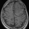
 Lobar Pneumonia. Chest radiograph of a lobar pneumonia, affecting the right middle lobe. Mikael Häggström, M.D. - Not altered. CC0

Bronchopneumonia
Bronchopneumonia is characterized by patchy, sporadic consolidation, concentrated on the bronchioles.
Bronchopneumonia is frequently bilateral and multifocal.
Bronchopneumonia is caused by different bacterial species.
 Bronchopneumonia. Histopathology of bronchopneumonia, showing neutrophils filling a bronchiole. Mikael Häggström, M.D. - CC0
Interstitial Pneumonia
Interstitial pneumonia is also known as atypical pneumonia.
Diffuse interstitial infiltrates are the defining features of interstitial pneumonia.
Interstitial pneumonia presents with only a few minor upper respiratory symptoms (low fever, little sputum) which is an ‘atypical’ presentation.
Interstitial pneumonia is typically caused by bacteria or viruses.
 Interstitial Pneumonia. Micrograph of usual interstitial pneumonia (UIP). UIP is the most common pattern of idiopathic interstitial pneumonia (a type of interstitial lung disease) and usually represents idiopathic pulmonary fibrosis. H&E stain. Autopsy specimen. No machine-readable author provided. KGH assumed (based on copyright claims). Not altered. CC BY-SA 3.0


Aspiration Pneumonia
Aspiration pneumonia is seen in individuals who are at risk for aspiration, such as alcoholics and people who are unconscious.
The oropharynx’s anaerobic bacteria, such as Bacteroides, Fusobacterium, and Peptococcus, are typical causes of aspiration pneumonia.
Aspiration pneumonia classically leads to a right lower lobe abscess (due to the angle of the bronchus), but may present in different lobes.
Compared to the left, the right main stem bronchus branches anatomically at a less sharp angle.
 Aspiration Pneumonia. Not altered. KGH CC BY-SA 3.0

Tuberculosis (TB)
As a result of first-time exposure to aerosolized Mycobacterium tuberculosis, primary tuberculosis (TB) develops.
Tuberculosis (TB) results in the hilar lymph nodes undergoing fibrosis and calcification to produce a Ghon complex as well as localized, caseating necrosis of the lung.
Although primary tuberculosis (TB) usually has no symptoms, it results in a positive purified protein derivative (PPD) test result.
When Mycobacterium tuberculosis reactivates, secondary tuberculosis (TB) develops.
Reactivation of Mycobacterium tuberculosis is frequently caused by AIDS, although it can also be caused other immune compromising issues such as disease or advanced age.
Tuberculosis (TB) usually occurs at the lung’s apex due to the high oxygen tension in that region of the lungs.
Tuberculosis (TB) creates a cavitary foci of caseous necrosis and has the potential to cause tuberculous bronchopneumonia or miliary pulmonary tuberculosis.
Symptoms of tuberculosis (TB) include:
- Fever
- Weight loss
- Night sweats
- Cough with hemoptysis
Histology of tuberculosis (TB) shows caseating granulomas, and positive acid-fast bacilli (AFB) stain.
Common sites for systemic dissemination of tuberculosis (TB) include:
- Meninges (meningitis)
- Cervical lymph nodes
- Kidneys (sterile pyuria)
- Lumbar vertebrae (Pott disease)
 Tuberculosis. Microscopy of tuberculous epididymitis. H&E stain Department of Pathology, Calicut Medical college. Not altered. CC BY-SA 4.0