Oral cavity pathology includes:
- Cleft lip
- Cleft palate
- Aphthous ulcer
- Behçet syndrome
- Oral herpes
- Squamous cell carcinoma
- Leukoplakia
- Oral candidiasis
- Hairy leukoplakia
- Erythroplakia
Cleft Lip
The five facial prominences grow and usually unite to create the face during the early stages of pregnancy.
The five facial prominences include:
- One superior
- One left
- One right
- Two inferiors
A cleft lip is a full thickness lip deformity brought on by the failure of the facial prominences to fuse together.
Cleft palate frequently coexists with cleft lip.
 Mild left unilateral cleft lip repair(A) Mild left unilateral cleft lip. (B) Four-week postoperative follow-up with good results. (C) Follow-up after six months. Outcomes of Primary Unilateral Cheiloplasty in Same-Day Surgical Settings. Khan M, Ullah H, Aziz A, Tahir M - Archives of plastic surgery (2016). Not altered. CC.


Cleft Palate
A complete thickness deformity in the palate known as cleft palate results from the inability of the facial prominences to fuse.
Cleft lip frequently coexists with cleft palate.
A single cleft palate is less frequent.
 Cleft Lip and Cleft Palate Malformation courtesy of Clapa Community

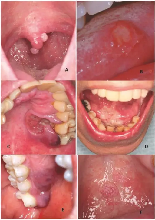
Aphthous Ulcer
A painful, superficial ulceration of the oral mucosa is known as an aphthous ulcer.
Aphthous ulcers develop as a result of physical strain, trauma, and friction.
Typically, aphthous ulcers have a gray foundation encircled by blush red erythema.
 Aphthous Ulcer. Not altered. CC BY-SA 3.0


Behçet Syndrome
Behçet syndrome includes:
- Uveitis
- Genital ulcers
- Recurrent aphthous ulcers
An immune complex vasculitis of the tiny vessels is what causes Behçet syndrome.
Behçet syndrome can cause recurrent aphthous ulcers.
 Behcet Syndrome. Claire Lewis. Not altered. CC.


Oral Herpes
Lesions in the oral cavity that are painful, red, and ulcerative can result from oral herpes.
HSV-1 is the most common cause of oral herpes.
HSV-2 can also cause oral herpes.
The initial oral herpes infection typically strikes children and preteens.
The herpes virus remains dormant in the trigeminal nerve ganglia after the initial sores have healed.
Stressors like tension, heat, sunlight, etc. can make the herpes virus reactivate.
The red pimples and ulcers return when the virus is reactivated.
 Oral Herpes. Herpetic gingivostomatitis. Note ulcers just below the front bottom teeth within the circle. James Heilman, Not altered. CC BY-SA 3.0


Squamous Cell Carcinoma
Squamous cell carcinoma (SCC) is the second most frequently diagnosed cancer worldwide.
Squamous cell carcinoma (SCC) accounts for about 30% of cancer cases in men and 20% of cases in women.
Two important risk factors for squamous cell carcinoma (SCC) are alcohol and tobacco use.
Invasive squamous cell carcinoma (SCC) may develop from oral leukoplakias and erythroplakias, which are precancerous diseases.
Although they can appear anywhere in the oral cavity epithelium, these lesions typically grow at the mouth’s floor.
Histologically, squamous cell carcinoma (SCC) is identified by alterations in the stratified squamous epithelial layers, such as invasion into the underlying connective tissue and hyperkeratosis, parakeratosis, acanthosis, keratin pearls, and dysplasia.
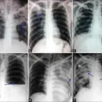
 Squamous Cell Carcinoma. A large head and neck squamous-cell carcinoma of the tongue as seen on CT imaging James Heilman, MD. Not altered. CC BY-SA 4.0


Leukoplakia
A frequent premalignant lesion of the mucous membranes of the mouth, throat, larynx, esophagus, and genitalia is called leukoplakia, sometimes known as white patch disease.
A white spot on the tissue’s surface that cannot be removed is the hallmark of leukoplakia.
Usually, neither bleeding nor ulcers occur in leukoplakia.
Thrush (oral candidiasis) and leukoplakia are entirely different conditions.
Hairy leukoplakia should not be confused with leukoplakia.
 Leukoplakia. Leukoplakia on the side of tongue Aitor III - Not altered. Public Domain


Oral Candidiasis
Candida is a yeast organism that is often present in minute amounts in regular saliva, is a frequent cause of oral thrush infection.
Candida is harmless to healthy individuals.
Candida overgrowth can happen in people whose immune systems have been compromised by disease or drugs.
As a result, the tissues in the mouth become inflamed, and “thrush” (a whitish crust or coating on the tongue’s surface) develops.
Risk factors for oral candidiasis (“thrush”) include:
- Antibiotic use like penicillin or cephalosporin
- Cancer chemotherapy drugs like cyclophosphamide
- Steroids
- Radiation therapy
- HIV/AIDS medications
- Organ transplant recipients
- Diabetes
- Weakened immune systems
- Severe burn injuries
- Leukemia
Thrush can occasionally progress into the potentially dangerous disorder oropharyngeal candidiasis (OPC).
 Oral Candidiasis. Thrush in a child who had taken antibiotics. James Heilman, MD - Not altered. CC BY-SA 3.0


Hairy Leukoplakia
Hairy leukoplakia (HL) is a histological manifestation of HIV infection and is distinguished by parakeratosis of the oral mucosa, hyperkeratosis, keratinization of the epithelium, and numerous intracellular inclusion bodies made up of lipid vacuoles, concentric lamellar structures, and occasionally viral particles within macrophages.
Oral sores are very contagious and frequently lead to the development of systemic diseases.
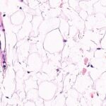
 Photomicrograph showing PAP-stained ground glass appearance of the nucleus and a peripheral nuclear bearding 100× magnification. Oral hairy leukoplakia: An exfoliative cytology study. Reginald A, Sivapathasundharam B - Contemporary clinical dentistry (2010). Not Altered. CC.
Erythroplakia
Erythroplakia is a red plaque that is a tissue lesion characterized by thickened epithelial mucosa with areas of white plaques.
Erythroplakia is also known as vascularized leukoplakias.
Red plaques are associated with oral cancer.
Erythroplakia usually occurs on the gingiva, buccal mucosa, lateral tongue surface, floor of mouth or retromolar trigone.
These lesions are most commonly found on the anterior surface of the maxillary alveolar ridge, but they have been reported to arise on the hard palate, vestibular sulcus and soft palate.
 Erythroplakia is defined as a red patch that cannot be diagnosed as any other lesion clinically and pathologically. Epithelial dysplasia in oral cavity. Shirani S, Kargahi N, Razavi SM, Homayoni S - Iranian journal of medical sciences (2014). Not Altered. CC.