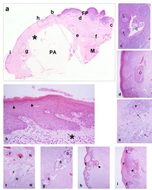
(a) Scanning view of a skin flap (asterisk) that has been transferred adjacent to lingual foliate papillae (FP). The panniculus adiposis (subcutaneous fat, PA), submucosal skeletal muscle (M), and the mammillated surface of the papillae are easily seen. The linear segment indicates the junction between the flap and native mucosa. (b) The linear segment indicates the junction between epidermis (left part of the photomicrograph) and the oral epithelium (right part of the photomicrograph). The stratum granulosum (arrowheads) of the epidermis is discernible. The lower border of both the epidermis and oral epithelium is irregular. This indicates hyperplasia of rete in response to the presence of non-specific chronic inflammation (asterisk) in underlying stroma. (c) Cross-sectioned profile of a crypt (C) of the lingual tonsil, which is surrounded by hyperplastic lymphoid tissue with germinal centers (arrows). Emigrant lymphoid cells and debris are present in the crypt lumen. (d) Cross-sectioned profile of a metaplastic collecting duct of the posterior superficial lingual salivary gland (Weber's). Lymphoid tissue with three germinal centers is present at the left of the photomicrograph. It is difficult to distinguish the surface oral epithelium at this magnification. (e) Two lobules of the posterior superficial lingual salivary gland, which are variably affected by parenchymal atrophy and non-specific chronic inflammation. Inflammation (asterisk) is prominent in the upper lobule, whereas mucous acini and tubules are preserved in the lower. The arrows indicate ectatic atrophic duct-like structures. (f) Randomly mixed fat (F), skeletal muscle (M), nerve fascicles (N), and vessels (V) characterize the submucosal soft tissues and contrast with the uniform and orderly arrangements of panniculus adiposis. (g) Two eccrine sweat-gland lobules. Paler secretory segments (S) and darker ducts (D) are distinguished. Adipocytes are mixed with the secretory segments. (h) Retiform downward growth of epidermis. The changes resemble senile (solar) lentigo or inchoate seborrhoeic keratosis. The structure indicated by the arrowhead is probably a cross-sectioned plugged infundibulum. (i) Area of ulceration covered by a fibrinous membrane (F). There is underlying non-specific chronic inflammation (asterisk). Histological changes in intra-oral skin flaps: Woolgar JA, Triantafyllou A - Head & neck oncology (2009). Not altered. CC.
Inflammation is the body’s response to injury or infection.
Inflammation is basically divided into two types:
Acute inflammation: Immediate.
Chronic inflammation: Long term.
 What are Causes of Inflammation?