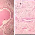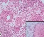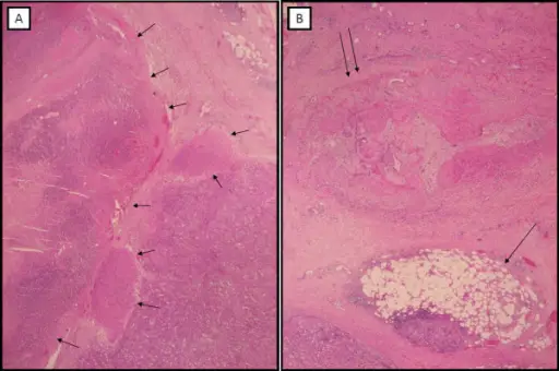
Fat necrosis is characterized by necrotic adipose tissue with foamy histiocytes, chronic inflammatory cells, and multinucleated giant cells.
What is the Pathology of Fat Necrosis?
The pathology of fat necrosis is:
-Etiology: Fat necrosis may be caused by trauma, iatrogenic injury, or radiation therapy.
-Pathogenesis: Fat necrosis.
-Morphology: The morphology associated with fat necrosis shows lipid laden histiocytes (lipophages), multinucleated giant cells, and degenerating adipocytes.
-Histology: The histology associated with fat necrosis shows necrosis of adipocytes.
How does Fat Necrosis Present?
Patients with fat necrosis typically are older females. The symptoms, features, and clinical findings associated with fat necrosis include a painless superficial solitary mass which may mimic cancer.
How is Fat Necrosis Diagnosed?
Fat necrosis can be diagnosed by physical exam, diagnosed by palpation of the breast mass, radiographical findings, and correlation with clinical history of trauma or breast procedures.
How is Fat Necrosis Treated?
Fat necrosis is treated by surgical excision, but it’s used only for painful or aesthetically undesirable lesions. Biopsy of the lesion is necessary to confirm the diagnosis.
What is the Prognosis of Fat Necrosis?
The prognosis of fat necrosis is good. It should resolve or regress over time, but the degenerated fat may persist for years within a fibrotic scar.