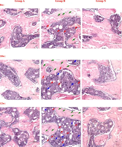
The observation of femoral head osteonecrosis by Hematoxylin-Eosin staining. In the group A, it showed partial necrotic changes in bone trabeculae and slight accumulation of degenerative or necrotic medullary haematopoietic cells and fat cells in the surrounding bone marrow. A few apparent empty lacunae were observed. The increase of fibroblasts and osteoclasts were not obvious. In the group B, the bone trabecular became sparse and fracture, more empty lacunae were seen in it. Haematopoietic cells and fat cells showed necrotic changes, the fibroblasts and osteoclasts increased and accumulated, and many activated osteoclasts were observed and resorbing the necrotic bone trabeculae. The performance of osteonecrosis became more and more serious with time. There was no visible necrosis of bone or bone marrow in the group N. (Magnification 200×, scale bar represents 100 μm for all figures, the green arrow stands for the empty lacunae, the red arrow stands for the multinucleated giant cells or osteoclasts, and the blue arrow stands for the necrosis or fibrosis of bone marrow).Immune response associated with Toll-like receptor 4 signaling pathway leads to steroid-induced femoral head osteonecrosis.
Tian L, Wen Q, Dang X, You W, Fan L, Wang K - BMC musculoskeletal disorders (2014). Not Altered. CC.
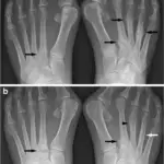
Osteonecrosis is a degenerative bone condition characterized by the death of cellular components of the bone secondary to an interruption of the subchondral blood supply.
What is the Pathology of Osteonecrosis?
The pathology of osteonecrosis is:
-Etiology: The causes of osteonecrosis are bone fractures, joint dislocations, alcoholism, and the use of high-dose steroids.
-Genes involved: ADH2, ADH3, ALDH2 and P450E1.
-Pathogenesis: The sequence of events that lead to osteonecrosis involves the death of cellular elements of the bone marrow.
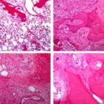
-Histology: The histology associated with osteonecrosis shows dead trabeculae stain deeper blue than non necrotic bone.
How does Osteonecrosis Present?
Patients with osteonecrosis typically affect both males and females 30-50 years of age. The symptoms, features, and clinical findings associated with osteonecrosis include pain and discomfort in a joint which increases over time.
How is Osteonecrosis Diagnosed?
Osteonecrosis is diagnosed by bone scintigraphy and MRI.
How is Osteonecrosis Treated?
Osteonecrosis is treated by NSAIDs, physiotherapy, and surgery.
What is the Prognosis of Osteonecrosis?
The prognosis of osteonecrosis is good. Most people fully recover from osteonecrosis and are eventually able to use the affected joint without pain.