What is Myocardial Infarction?

Myocardial Infarction
Myocardial Infarction. Example of a 76 year old male patient with an acute myocardial infarction (STEMI) of the anterior wall after acute PCI of the occluded LAD with stent implantation (pain-to-balloon time 150 min.). a The water sensitive T2-STIR image demonstrates the edema in the anterior wall (white arrows), c the LGE images demonstrate an almost transmural myocardial infarction (white arrows) with central MVO (asterisk). This indicates almost no myocardial salvage after successful revascularization of the LAD. b shows the T2* image at TE = 15 ms and d the T2* imaging map of the T2* mapping. The central dark area (white arrow) represents pixels with a T2* decay <20 ms indicating postreperfusionhemorrhage. Assessment of acute myocardial infarction: current status and recommendations from the North American society for Cardiovascular Imaging and the European Society of Cardiac Radiology. Stillman AE, Oudkerk M, Bluemke D, Bremerich J, Esteves FP, Garcia EV, Gutberlet M, Hundley WG, Jerosch-Herold M, Kuijpers D, Kwong RK, Nagel E, Lerakis S, Oshinski J, Paul JF, Underwood R, Wintersperger BJ, Rees MR, North American Society of Cardiovascular. ImagingEuropean Society of Cardiac Radiolo - The international journal of cardiovascular imaging (2010). Not Altered. CC.

Myocardial infarction is an irreversible heart muscle death caused by ischemia.

What is the Pathology of Myocardial Infarction?

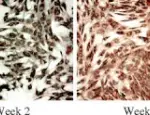
Myocardial infarction pathology is characterized by death of heart muscle (necrosis) due to blocked arteries.

How does Myocardial Infarction Present?

Myocardial infarction affects millions of Americans annually. Individuals typically present with chest pain that is preceded by fatigue, chest discomfort and malaise. Chest pain is prolonged and continuous for 30-60 minutes with radiation to neck, shoulder, jaws and left arm. 

How is Myocardial Infarction Diagnosed?

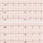
Myocardial infarction diagnosis include electrocardiogram, cardiac markers, CBC, metabolic panel, and lipid profile. Usual electrocardiogram findings are: ST-segment elevation, presence of new Q waves, ST-segment depression, T wave inversion. And nonspecific ST-T wave abnormalities.

How is Myocardial Infarction Treated?

Myocardial infarction treatment includes restoration of perfusion with percutaneous coronary intervention (PCI) or coronary artery bypass graft (CABG). Other pharmacologic management include: statins, aspirin, nitrates, and analgesia.

What is the Prognosis of Myocardial Infarction?

Myocardial infarction prognosis is poor with associated mortality of 30%; and 10% of survivors die after 1 year.