Pathologic calcification is the deposition of calcium salts, typically as phosphates or carbonates, in soft tissues (i.e., tissues that would not be calcified in a healthy state). If calcification is extensive, it appears grossly as chalky white deposits with a brittle or gritty texture. There are two different types of pathologic calcification: dystrophic calcification and metastatic calcification.
What is Dystrophic Calcification?
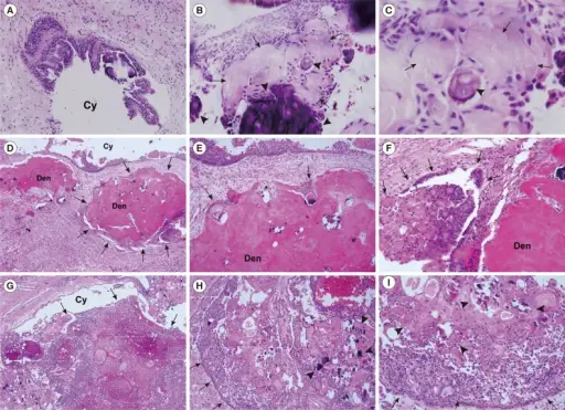
Dystrophic calcification is the calcification of dead tissue as part of the process of necrosis. Calcification is the gross lesion for which the myocardial and skeletal muscle necrosis of vitamin E or selenium deficiency in ruminants was named white muscle disease. Calcification is also prominent in other forms of necrosis (e.g., in the caseous necrosis of tuberculoid granulomas), in parasitic granulomas, and in necrotic fat or lipomas (benign neoplasms of adipocytes).
What is Metastatic Calcification?
Metastatic calcification is soft tissue calcification as the result of elevated serum calcium concentration. It targets the intima and tunica media of vessels, especially those in the lungs, pleura, endocardium, kidneys, and stomach. The primary defect is an imbalance in calcium and phosphate concentrations in the blood.


