Neuronal inclusions are basophilic round, oval or crescentic intracytoplasmic inclusions that stain pale blue with H&E staining. Occasionally small vacuoles are found within neuronal inclusions. Neuronal inclusions can be the size of the nucleus of a neuron.
What are Neuronal Inclusions?
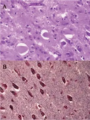
Pathological findings of the cingulate gyrus and the amygdale of the dominant hemisphere. (A) Hematoxylin-eosin staining reveals many ballooned neurons (Pick cells) in the left cingulate gyrus. (B) Bodian staining shows many argyrophilic neuronal inclusions (Pick bodies) in the left amygdala.Primary progressive apraxia of speech (AOS) in a patient with Pick's disease with Pick bodies: a neuropsychological and anatomical study and review of literatures.
Uyama N, Yokochi F, Bandoh M, Mizutani T - Neurocase (2012). Not Altered. CC.
Post navigation
Previous Post
 What is Axonal Reaction?
Next Post
How do Astrocytes React to Injury?