Syringomyelia is the development of a fluid-filled cyst (syrinx) within the spinal cord. Over time, the cyst can enlarge and destroy the spinal cord.
What is the Pathology of Syringomyelia?
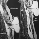
Etiology: The causes of syringomyelia may include Chiari malformation, infections, and trauma.
Pathogenesis: The sequence of events that lead to Syringomyelia is the formation of a syrinx. When there is increased cerebrospinal fluid contained within the ependyma of the central canal of the spinal cord, it dissects the surrounding white matter to form this cystic cavity. A number of pathological conditions can cause an obstruction of the normal cerebrospinal fluid spaces, as discussed above.
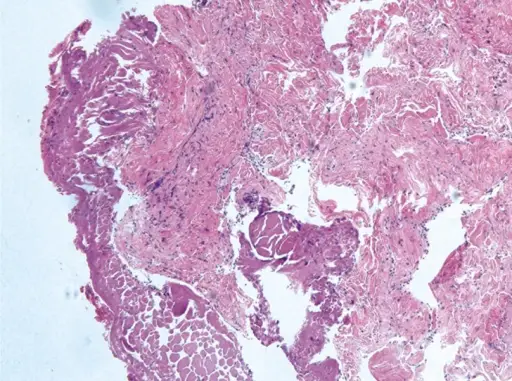
Histology: The histology associated with syringomyelia shows cavitation of spinal cord gray matter and syrinx adjacent to the central canal.
How does Syringomyelia Present?
Patients with syringomyelia typically present between 20 and 40 years old, and men are more affected than women. The symptoms, features, and clinical findings associated with syringomyelia include muscle weakness, headaches, and stiffness.
How is Syringomyelia Diagnosed?
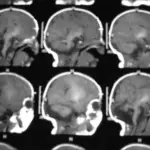
Syringomyelia is diagnosed via MRI.
How is Syringomyelia Treated?
Syringomyelia is treated with surgery.
What is the Prognosis of Syringomyelia?
The prognosis of syringomyelia depends on the underlying cause.