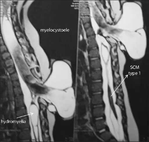
Hydromyelia is dilatation of the central canal of the spinal cord.
What is the Pathology of Hydromyelia?
Etiology: The causes of hydromyelia includes infections, spinal tumors, and trauma.
Pathogenesis: The sequence of events that lead to hydromyelia is due to the abnormal widening of the central canal of the spinal cord that allows the buildup of fluid.
How does Hydromyelia Present?
Patients with hydromyelia typically are males rather than females with an age range of 25-40 years. The symptoms, features, and clinical findings associated with hydromyelia include loss of feeling in the hands and arms, leg pain, and muscle weakness.
How is Hydromyelia Diagnosed?
Hydromyelia is diagnosed with MRI. Additional tests include electromyography, CT scan, and myelogram.
How is Hydromyelia Treated?
Hydromyelia is treated by surgery and physical therapy.
What is the Prognosis of Hydromyelia?
The prognosis of hydromyelia is good and it may rarely resolve on its own without medical intervention. Surgery may permanently or temporarily relieve symptoms, but it can also cause a number of severe complications.