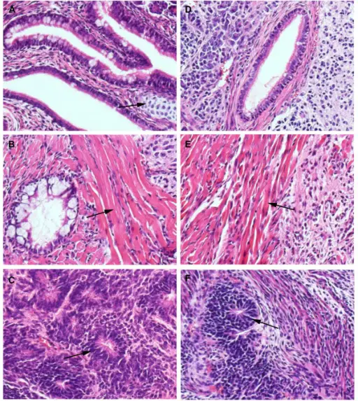
Teratomas are rare tumors that may hold different types of tissue such as bone, teeth, muscle, and hair. Teratomas are mostly found in the ovaries, testicles, and sacrum (tailbone), but also sometimes grow in the nervous system and abdomen. A teratoma may be cancerous or benign.
What is the Pathology of Teratomas?
The pathology of teratomas is:
–Etiology: The cause of teratomas is due to abberrent cellular differentiation.
-Genes involved: Unknown.
-Pathogenesis: The sequence of events that lead to teratomas is a pathological transformation of primordial germ cell during cellular development.
–Morphologic changes: The morphologic changes involved with (teratomas):
How does Teratomas Present?
Patients with teratomas are typically females, but it can happen in males. Teratomas most commonly occur in the tailbone, ovaries, and testicles, but can occur elsewhere in the body. Teratomas can appear in newborns, children, or adults. The symptoms, features, and clinical findings associated with teratomas include: growing masses, swelling, constipation, incontinence and leg weakness.
How are Teratomas Diagnosed?
Teratomas are diagnosed by blood tests that test for elevated levels of the alpha fetoprotein (AFP) and beta human chorionic gonadotrophin hormone (BhCG). Ultrasound imaging can help identify the progress of the teratoma. To check if cancer has spread to other parts of the body, your doctor will request X-rays of your chest and abdomen. Blood tests are also used to check for tumor markers.
How are Teratomas Treated?
Teratomas are treated by surgical removal of tumors or involved organs, performed by a pediatric surgeon, and with chemotherapy if the teratoma is malignant.
What is the Prognosis of Teratomas?
The prognosis of teratomas is not good. Older age at diagnosis, higher stage and grade confer worse prognosis but overall improved after chemotherapy, with approximately 75% survival for stage IV.


