Wilms tumor is is a type of childhood cancer that starts in the kidneys. It is the most common type of kidney cancer in children. It is also known as nephroblastoma.
What is the Pathology of Wilms Tumor?
The pathology of Wilms tumor is:
–Etiology: The cause of Wilms tumor is alterations of genes responsible for normal genitourinary development.
-Genes involved: Mutations in the WT1 gene, CTNNB1 gene, or AMER1 gene.
–Pathogenesis: The sequence of events that lead to Wilms tumor are malignant growth of primative cells containing metanephric blastema, stromal and epithelial derivatives. Dysfunction is caused when the tumor compresses the normal kidney parenchyma
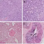
–Morphologic changes: The morphologic changes involved with Wilms tumor are mixed pattern with blastema, stroma and single epithelial structures. Characteristic features of Wilms tumor are the presence of abortive tubules and glomeruli surrounded by a spindled cell stroma. The stroma may include striated muscle, cartilage, bone, fat tissue, and fibrous tissue.
How does Wilms Tumor Present?
Patients with Wilms tumor are typically females between 2 to 8 years old. The symptoms, features, and clinical findings associated with Wilms tumor include abdominal swelling, abdominal pain, and perhaps an abdominal mass.
How is Wilms Tumor Diagnosed?
Wilms tumor is diagnosed by urinalysis, CT scan, X-rays to check size of tumor. Biopsy can confirm the diagnosis.
How is Wilms Tumor Treated?
Wilms tumor is treated by surgical removal followed by radiotherapy or chemotherapy.
What is the Prognosis of Wilms Tumor?
The prognosis of Wilms tumor is good. More than 80% of all children diagnosed with Wilms tumor are expected to survive the disease long-term. With a timely diagnosis before the tumor has metastasized to other parts of the body the cure rate is even higher at 90% with standard treatment.