Cholesteatomas are skin-lined cysts in the ear.
What is the Pathology of Cholesteatomas?
The pathology of cholesteatomas is:
-Etiology: The cause of cholesteatomas is a chronic ear infection, colds, sinus infections, allergies, or an abnormal functioning eustachian tube.
-Pathogenesis: The sequence of events that lead to cholesteatomas is an infection in the middle ear which may affect the eustachian tube. If the eustachian tube is not working correctly, pressure build up within the middle ear can pull part of the eardrum the wrong way. This pulling of the eardrum may create a cyst or sac that fills with skin cells.
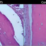
-Histology: The histology associated with cholesteatomas shows keratinized stratified squamous epithelium with granulation tissue. A chronic inflammatory infiltrate and keratin debris may also be appreciated.
How do Cholesteatomas Present?
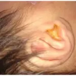
Patients with cholesteatomas typically are males in their 20s or 30s. The symptoms, features, and clinical findings associated with cholesteatomas include a feeling of pressure, a bump in the ear, and possibly ear drainage. Cholesteatomas are usually painless.
How are Cholesteatomas Diagnosed?
Cholesteatomas are diagnosed by a physical exam to look for a visible deposit of skin cells or a large mass in the ear.
How are Cholesteatomas Treated?
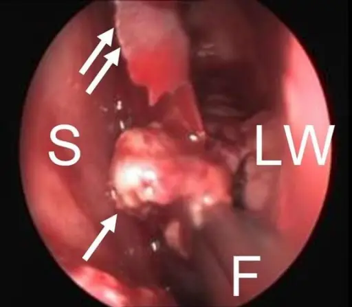
Cholesteatomas are treated with a surgical treatment.
What is the Prognosis of Cholesteatomas?
The prognosis of cholesteatomas is fair.