Celiac disease is a digestive and autoimmune disorder that can damage your small intestine. Celiac disease can be triggered by a protein called gluten. Gluten is found in grains, like wheat, barley and rye.
What is the Pathology of Celiac Disease?
The pathology of celiac disease is:
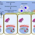
-Etiology: The cause of celiac disease is by sensitivity to the gliadin fraction of gluten. In a genetically susceptible person, gluten-sensitive T cells are activated when gluten-derived peptide epitopes are presented.
-Genes involved: HLA-DQA1 and HLA-DQB1 genes.
-Pathogenesis: The sequence of events that lead to celiac disease are: activation of both a cell-mediated T-cell and humoral B-cell immune response resulted from exposure to the glutens. Transglutaminase deamidates gliadin which results in greater proliferative response of gliadin-specific T-cells that lead to mucosal inflammation and further B-cell activation in patients with the disease.
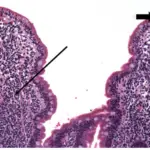
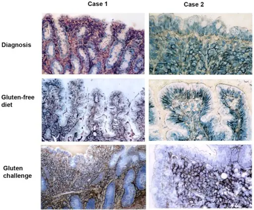
-Histology: The histology associated with celiac disease shows intraepithelial lymphocytosis, lamina propria inflammation and villous atrophy.
How does Celiac Disease Present?
Patients with celiac disease typically in women at any age from infancy well into senior adulthood. The symptoms, features, and clinical findings associated with celiac disease include diarrhea, fatigue, weight loss, bloating and gas, abdominal pain, nausea and vomiting, and constipation.
How is Celiac Disease Diagnosed?
Celiac disease is diagnosed by serology testing looks for antibodies, Genetic testing for human leukocyte antigens, intestinal biopsy.
How is Celiac Disease Treated?
Celiac disease is treated by following a gluten-free diet.
What is the Prognosis of Celiac Disease?
The prognosis of celiac disease is fair. Overall, people with untreated or unresponsive celiac disease have increased early mortality compared to the general population.