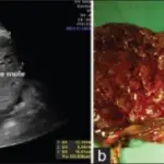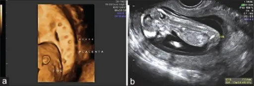
A partial mole is a mole where there might be identifiable fetal residues.
What is the Pathology of Partial Mole?
The pathology of the partial mole is:
-Etiology: The cause of partial mole is excess of paternal chromosomes promotes the growth of the placental tissue.
-Genes involved: None.
-Pathogenesis: The sequence of events that lead to partial mole shows triploid, (69 XXY, 69, XXY, 69, XXX), abnormal fetus/embryo, focal swelling of villi, focal trophoblastic hyperplasia.
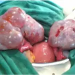
-Morphology: The morphology associated with partial mole shows villi are vesicular, whereas others appear more normal, and embryonic/fetal development may be seen but the fetus is always malformed and is never viable.
-Histology: The histology associated with partial mole shows heterogeneity in villous size with 2 discrete populations; large, hydropic villi, and small, fibrotic villi.
How does Partial Mole Present?
Patients with partial mole typically affect female present at the age range of 20-55 years. The symptoms, features, and clinical findings associated with partial mole include painless vaginal bleeding, the uterus may be larger than expected, or the ovaries may be enlarged, hyperemesis, increased blood pressure, high HCG levels.
How is Partial Mole Diagnosed?
Partial mole is diagnosed using blood tests and histopathological examination.
How is Partial Mole Treated?
Partial mole is treated uterine suction or by surgical curettage, and methotrexate.
What is the Prognosis of Partial Mole?
The prognosis of the partial mole is good if treated in a timely manner.