Pleural effusions are an accumulation of excess fluid in the pleura cavity of the lungs.
Types of Pleural Effusion: Inflammatory Pleural Effusions, Noninflammatory Pleural Effusions.
What is the Pathology of Pleural Effusion?
The pathology of pleural effusion is the gross and microscopic study of the factors like heart failure that lead to the accumulation of fluid in the pleura and how to manage them.
-Etiology: The cause of the pleural effusion is mostly congestive heart disease, pulmonary embolism, and pneumonia.
-Genes involved: None.
-Pathogenesis: The sequence of events that lead to pleural effusion are pleural inflammation altering the permeability of the membranes with impaired lymphatic drainage in the pleural space that leads to accumulation of excess fluid which can be in form of exudate or transudates.
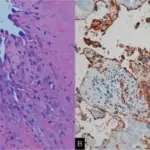
-Histology: The histology associated with pleural effusion shows clear layers of simple cuboidal mesothelium cells found on top of connective tissues.
How does Pleural Effusion Present?
Patients with pleural effusion are typically male, female, and children with an age range of determining age. The symptoms, features, and clinical findings associated with pleural effusion include chest pain, shortness of breath, cyanosis, cough, edema, and orthopnea.
How is Pleural Effusion Diagnosed?
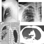
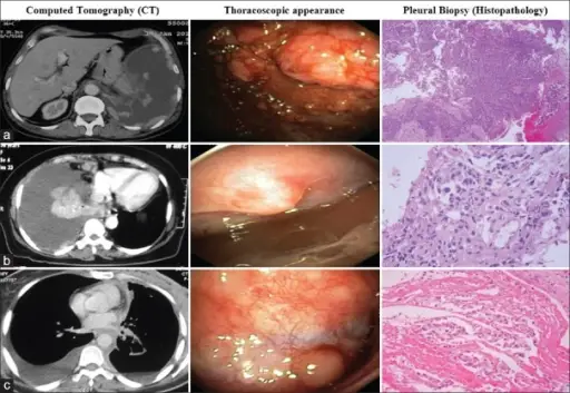
Pleural effusion is diagnosed mostly by use of CT scan since it clearly shows the area with the excess fluid. Other ways include a physical exam and history taking.
How is Pleural Effusion Treated?
A patient with pleural effusion is treated through a pleural tap, therapeutic thoracocentesis, tube thoracostomy, and pleurodesis to remove the excess fluid.
What is the Prognosis of Pleural Effusion?
The prognosis of pleural effusion is poor in the malignant form since it has a survival rate of only 4 months.