Ganglion cysts are nodular lesions that most commonly develop in the wrist.
What is the Pathology of Ganglion Cysts?
The pathology of ganglion cysts is:
-Etiology: The cause of ganglion cysts is unknown.
-Genes involved: None.
-Pathogenesis: The sequence of events that lead to ganglion cysts is unknown.
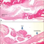
-Histology: The histology associated with ganglion cysts shows cystic lesions that are lined by connective tissue. Ganglion cysts are typically composed of bland spindle-shaped cells in a background of myxoid matrix.
How does Ganglion Cysts Present?
Patients with ganglion cysts typically present with pain, and a subcutaneous nodule.
How are Ganglion Cysts Diagnosed?
Ganglion cysts are usually diagnosed utilizing physical examination, ultrasound, and needle aspiration.
How Are Ganglion Cysts Treated?
Many ganglion cysts resolve on their own. Needle aspiration or surgery may be considered for certain patients.
What is the Prognosis of Ganglion Cysts?
The prognosis of ganglion cysts is good.