Horseshoe kidney is a congenital condition characterized by the fusion of the upper or lower poles of the kidneys.
What is the Pathology of Horseshoe Kidney?
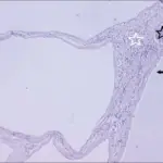
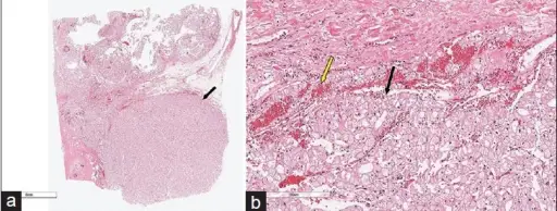
The pathology of horseshoe kidney is:
-Etiology: The cause of horseshoe kidney is idiopathic, teratogenic event,
-Genes involved: Unknown.
-Pathogenesis: The sequence of events that lead to horseshoe kidney not understood.
-Morphology: The morphology associated with horseshoe kidney shows fused kidneys.
How does Horseshoe Kidney Present?
Patients with horseshoe kidney typically twice more common in males than females present at age range prenatal. The symptoms, features, and clinical findings associated with horseshoe kidney include asymptomatic, frequent UTI, abdominal pain, nausea, abdominal swelling and fullness.
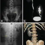
How is Horseshoe Kidney Diagnosed?
Horseshoe kidney is diagnosed through laboratory studies such asurinalysis, and serum chemistry with creatinine for baseline renal function. Imaging studies such as CT scanning of the abdomen and pelvis. Voiding cystourethrography rules out vesicoureteral reflux.
How is Horseshoe Kidney Treated?
Horseshoe kidney is treated through symptomatic and supportive care. Surgical care therapy.
What is the Prognosis of Horseshoe Kidney?
The prognosis of horseshoe kidney is good. Presence of the condition alone doesn’t affect survival.