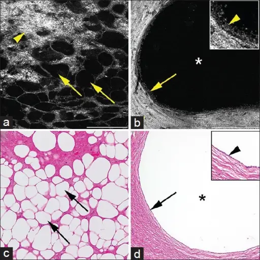
Full-field optical coherence tomography (a and b) and corresponding H&E images of (c and d) of benign kidney tumors. (a) Angiomyolipoma showing signal void polygonal adipocytes (arrows) and bright connective tissue from collagen (arrowhead). (b) Cystic nephroma showing large signal void cyst (*) lined by single-layered epithelium with dull gray signal (arrow and inset with arrowhead) embedded in thick collagenous tissue (bright signal; arrowhead). Full-field optical coherence tomography; scale bars (a) = 0.25 mm, (b) = 0.5 mm. Inset 2 × zoom of images B. H&E (c and d); total magnifications = ×200. Rapid evaluation of fresh ex vivo kidney tissue with full-field optical coherence tomography: Jain M, Robinson BD, Salamoon B, Thouvenin O, Boccara C, Mukherjee S - Journal of pathology informatics (2015). Not altered. CC.
Benign kidney neoplasms are renal tumor lesions that are primarily located in the kidney without the capability to invade other sites of the body.
Examples of benign kidney neoplasms include:
- Angiomyolipoma
- Oncocytoma
- Renal papillary adenoma
