Pineal gland pathology refers to any condition that may damage or alter the structure or function of the pineal gland.
WHAT IS PINEAL GLAND PATHOLOGY?
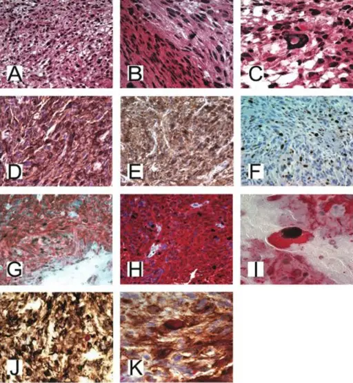
Immunohistochemical slides of malignant papillary glioneuronal tumor of the pineal gland. (A) Hematoxylin and Eosin staining at 20× magnification showing glial processes. (B) Hematoxylin and Eosin Stain 20× showing glial architecture. (C) Hematoxylin and Eosin staining at 40× showing abnormal nuclei pattern. (D and E) Synaptophysin positive indicating the neuronal nature of the tumor. (F) Ki67 proliferative index is strongly positive at 20%. (G and H) GFAP and Ki67 staining at 20× showing the strong proliferative index of the abnormal glial cells. (I) GFAP and Ki67 immunostaining at 100× demonstrating the strong proliferative index of the abnormal glial cell. (J and K) MAP and Ki67 combined immunostaining demonstrating the high proliferative indices within the abnormal neuronal cells. Malignant papillary glioneuronal tumor of the pineal gland: Case presentation and literature review of a distinct entity: Kaloostian PE, Chen H, Tran HP - The American journal of case reports (2013). Not altered. CC.
Post navigation
Previous Post
 What are Metastasis to the Liver?
Next Post
What are Pinealomas?