Pineocytomas are rare pineal parenchymal tumor that is well differentiated.
What is the Pathology of Pineocytomas?
The pathology of pineocytomas is:
-Etiology: The cause of pineocytomas is unknown. No specific genetic alterations and also no identified risk factors have been found regarding pineocytomas.
-Genes involved: Unknown.
-Pathogenesis: Aberrant cellular proliferation.
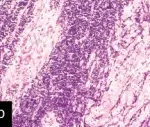
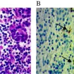
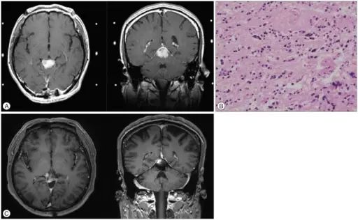
-Histology: Pinealcytomas is made up of cells in a uniform pattern that may form large pineocytomatous rosettes, pleomorphic cells with gangliocytic differentiation, or both.
How do Pineocytomas Present?
Patients with pineocytomas are typically females between 20 and 60 years old. Signs and symptoms include headache, ataxia, nausea, impaired vision, and loss of upward gaze.
How are Pineocytoma Diagnosed?
Pineocytoma is diagnosed by CT scanning, MRI and CSF. Confirmation of diagnosis is via brain biopsy.
How is Pineocytoma Treated?
Pineocytoma are treated by surgical excision. Some patients undergo chemotherapy or radiation therapy, determined individually per patient.
What is the Prognosis of Pineocytoma?
The prognosis of pineocytoma is good. There is low recurrence rates with gross total resection.