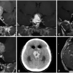
Hyperpituitarism is a clinical manifestation of the pathology of the pituitary gland characterized by spare secretion of trophic hormones, hyperfunction of the anterior pituitary gland.
What is Hyperpituitarism?
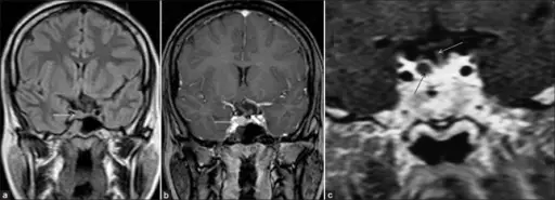
Pituitary microadenoma: Coronal FLAIR (fluid-attenuated inversion recovery) (a) and routine T1-weighted postcontrast (b) images of the brain show a small nodular lesion (thin white arrow) involving the right side of the pituitary gland producing a mild bulge of the superior margin of the gland and leftward deviation of the pituitary stalk (thick white arrow). The lesion appears isointense to the gland on the FLAIR image and shows an enhancement pattern almost identical to the normal pituitary gland. High resolution dynamic contrast-enhanced T1-weighted coronal image (c) of the brain of another patient (at 60 seconds) shows a small non-enhancing (dark) microadenoma (thin black arrow) lateralized to the right side of the pituitary gland. Note that the lesion is more conspicuous on dynamic contrast scan as compared with routine contrast scan (seen in Figure 5b). The normal pituitary gland shows marked homogenous enhancement and there is no deviation of the pituitary stalk (thin white arrow) in this case.
Post navigation
Previous Post
 What are the Cells of the Pituitary Gland?