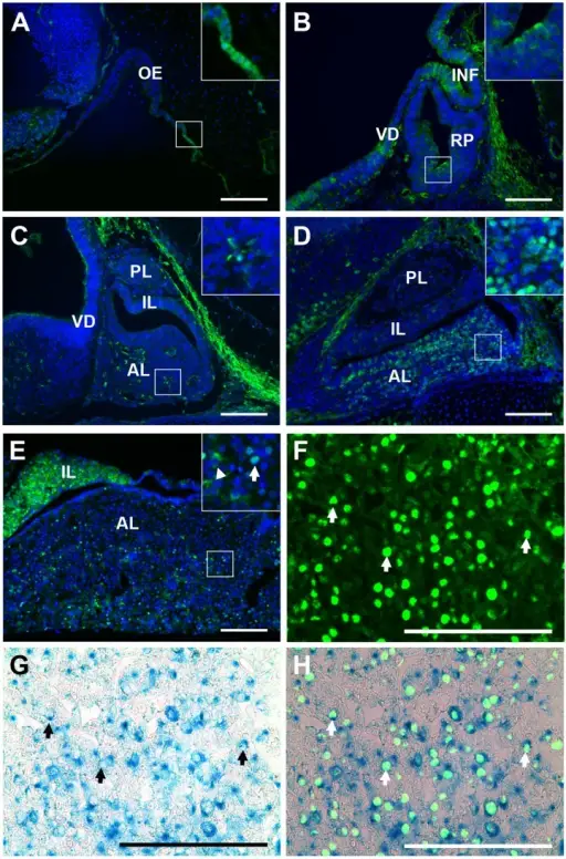
FOXO1 is present in the nuclei of pituitary cells at an increasing frequency as development progresses. Immunohistochemistry for FOXO1 (green) was performed on midsagittal pituitary sections. (A) FOXO1 is present in the developing pituitary by e10.5. Nuclear FOXO1 is apparent where the invaginating Rathke’s pouch is joined to the oral ectoderm that will form the mouth (see inset). (B) By e12.5 FOXO1 is present almost entirely in the cytoplasm of pituitary cells. (C) A few pituitary cells contain nuclear FOXO1 at e14.5. (D) At e18.5 the developing pituitary contains a mostly nuclear FOXO1. (E) In adults, FOXO1 is present in the anterior and intermediate lobes of the pituitary gland, but not in the posterior lobe (data not shown). In the adult pituitary FOXO1 is primarily nuclear (inset, arrow). Some cytoplasmic FOXO1 (inset, arrow head) is also present. (F) Immunohistochemistry for FOXO1. (G) Beta-galactosidase staining of pituitary from Foxo1+/LacZ mice identifies cells in which the endogenous Foxo1 promoter is active. (H) An overlay of immunohistochemical staining for FOXO1 (green) and β-galactosidase staining of pituitary from Foxo1+/LacZ mice (blue). (F–H) Arrows highlight examples of co-localized cells. Pictures are taken at 200X (A–E) or 630X (F–H). Insets are magnified 600X. Scale bars represent 100 µm. All cell nuclei were marked with DAPI (A–E, blue). Oral ectoderm (OE), infundibulum (INF), ventral diencephalon (VD), Rathke’s pouch (RP), posterior lobe (PL), intermediate lobe (IL), anterior lobe (AL). Forkhead Box O1 is present in quiescent pituitary cells during development and is increased in the absence of p27 Kip1: Majumdar S, Farris CL, Kabat BE, Jung DO, Ellsworth BS - PloS one (2012). Not altered. CC.
The anterior pituitary gland consists of acidophils, basophils, and chromophobe cell types.
Acidophils of the pituitary glands is a histological feature of the anterior pituitary, array of cells containing eosinophilic cytoplasm.
Basophils of the pituitary glands a histological feature of the anterior pituitary, array of cells containing basophil cytoplasm.
Chromophobes of the pituitary gland a histological feature of the anterior pituitary, array of cells containing poorly staining cytoplasm.
The six specific types of cells of the anterior pituitary gland include:
Regulates the secretion of glucocorticoids by the adrenal cortex
Gonadotrophs
Follicle Stimulating Hormone (FSH), and luteinizing hormone (LH)
In men, FSH stimulates spermatogenesisand LH regulates Leydig cell function.In women, FSH is involved in the regulation of follicle growth,while LH is related to ovulation and development of the corpus luteum
Lactotrophs
Prolactin
This stimulates the breast during lactation
Mammosomatotrophs
Prolactin
This stimulates the breast during lactation
Somatotrophs
Growth Hormone (GH)
Direct effects, the stimulation of protein synthesis in liver and muscle and lipolysis of fat stores. Indirect effects, skeletal growth mediated by insulin-like growth factor I (IGF-I), which also exerts negative feedback on GH release from the pituitary