Testicular atrophy is a regressive alteration affecting the scrotal testis, shrinking of the testis.
What is the Pathology of Testicular Atrophy?
The pathology of testicular atrophy is the gross and microscopic study of the structure and functional changes in testicular atrophy.
-Etiology: The cause of testicular atrophy is multifactorial including hypopituitarism, atherosclerotic tapering of the blood supply, cachexia, exhaustion atrophy, orchitis.
-Genes involved: Unknown.
-Pathogenesis: The sequence of events that lead to testicular atrophy are of multifactorial origin, inflammations, diet, underlying disease, congenital anomalies among others.
-Morphology: The morphology associated with testicular atrophy shows it is unilateral in most cases, bilateral in 25% of patients.
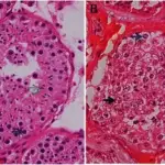
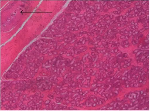
-Histology: The histology associated with testicular atrophy shows marked hyalinization and thickening of the basement membrane of the spermatic tubules.
How does Testicular Atrophy Present?
Patients with testicular atrophy typically affect males present at age range of 45 years. The symptoms, features, and clinical findings associated with testicular atrophy include smaller than normal testis, Leydig cell hyperplasia, history or presence of cryptorchidism.
How is Testicular Atrophy Diagnosed?
Testicular atrophy is diagnosed through physical examination. Imaging may be helpful.
How is Testicular Atrophy Treated?
Testicular atrophy is treated by hormonal therapy.
What is the Prognosis of Testicular Atrophy?
The prognosis of testicular atrophy is fair with proper management.