Pancreatic carcinoid tumor is a rare tumor of the pancreas that usually presents with carcinoid syndrome, which is characterized by diarrhea, valvular disease, cutaneous flushing.
What is the Pathology of Pancreatic Carcinoid Tumor?
The pathology of pancreatic carcinoid tumor is:
-Etiology: The cause of pancreatic carcinoid tumor is not known
-Genes involved: None.
-Pathogenesis: The sequence of events that lead to pancreatic carcinoid tumor are not known.
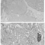
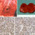
-Morphology: The morphology associated with pancreatic carcinoid tumor shows a polypoid mass.
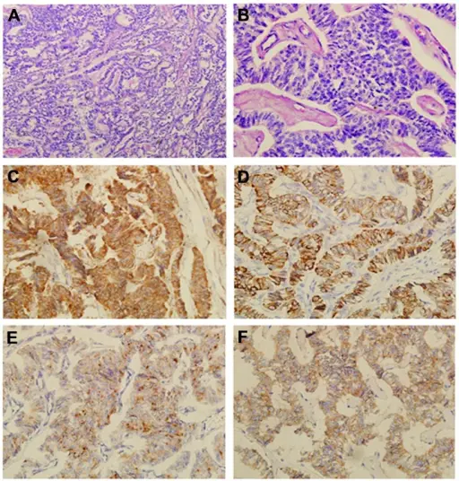
-Histology: The histology associated with pancreatic carcinoid tumor shows distinctive patterns: trabecular or ribbons; solid, nodular, and insular cords; ill differentiated or atypical forms; tubules and glands or rosettelike forms; and assorted patterns.
How does Pancreatic Carcinoid Tumor Present?
Patients with pancreatic carcinoid tumor typically have no gender prevalence present at an age range of 20 years and above. The symptoms, features, and clinical findings associated with pancreatic carcinoid tumor include abdominal pain, cutaneous flushing, and pellagra skin lesions.
How is Pancreatic Carcinoid Tumor Diagnosed?
Pancreatic carcinoid tumor is diagnosed through laboratory testing which includes biomarkers and biogenic amine levels. Imaging studies plain radiography, CT scan, ultrasound, MRI, and angiography.
How is Pancreatic Carcinoid Tumor Treated?
Pancreatic carcinoid tumor is treated through chemotherapy and surgical interventions.
What is the Prognosis of Pancreatic Carcinoid Tumor?
The prognosis of pancreatic carcinoid tumors is fair.