What is Varicella-Zoster Virus Infectious Neuropathy?

Varicella-Zoster Virus Infectious Neuropathy
Varicella-zoster virus (VZV) antigen in giant cell arteritis (GCA)–positive and GCA-negative temporal arteries (TAs) and inflammation adjacent to VZV antigen in GCA-negative TAsVZV antigen in TAs of GCA-positive and GCA-negative patients (clinical and laboratory features of GCA whose TA biopsies were pathologically negative for GCA) was detected immunohistochemically using mouse anti-VZV gE immunoglobulin (Ig) G1 antibody (Santa Cruz Biotechnology, Dallas, TX; catalog no. SC-56995). After immunostaining with primary and biotinylated secondary antibodies, slides were treated with prediluted streptavidin-alkaline phosphatase (BD Biosciences, San Diego, CA) for 1 hour. The color reaction was developed under a light microscope using the fresh fuchsin substrate system (Dako, Carpinteria, CA) with levamisole (Dako; 24 μg/mL). VZV antigen is shown in the adventitia of a positive control VZV-infected cadaveric cerebral artery 14 days after infection in vitro (A), in the adventitia and media of a GCA-positive TA (B), and in the media and intima of a GCA-negative TA (C). No staining was seen when mouse isotype IgG1 was substituted for primary mouse anti-VZV gE IgG1 antibody (D–F). In GCA-negative TAs, hematoxylin & eosin (H&E) staining of TA sections adjacent to those containing VZV antigen revealed inflammatory cells (G, J, arrows) identified as CD45-positive (H, K, pink) after destaining H&E sections and immunostaining with rabbit anti-CD45 antibody (Abcam, Cambridge, MA; catalog no. AB10558). No staining was seen when normal rabbit serum was substituted for rabbit anti-CD45 antibody (I, L). 600× magnification. VZV in biopsy-positive and -negative giant cell arteritis: Analysis of 100+ temporal arteries: Gilden D, White T, Khmeleva N, Boyer PJ, Nagel MA - Neurology® neuroimmunology & neuroinflammation (2016). Not altered. CC.

Varicella-Zoster virus infectious neuropathy is a common viral infection to the peripheral nervous symptoms.

What is the Pathology of Varicella-Zoster Virus Infectious Neuropathy?

The pathology of varicella-zoster virus infectious neuropathy is: 

-Etiology: The cause of varicella-zoster virus infectious neuropathy is Varicella-Zoster Virus Infectious complication.

-Genes involved: NA.

-Pathogenesis: The sequence of events that lead to varicella-zoster virus infectious neuropathy results from viral infections of the peripheral nervous system. Latent infection of neurons in the sensory ganglia of the spinal cord and brain stem follows chickenpox. Reactivation causes painful, vesicular skin outbreaks at the site of sensory dermatomes.

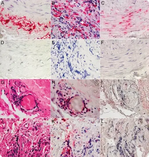
-Morphology: The morphology associated with varicella-zoster virus infectious neuropathy shows necrosis and hemorrhage at the site.

-Histology: The histology associated with varicella-zoster virus infectious neuropathy shows inflammatory insinuates of mononuclear. 

How does Varicella-Zoster Virus Infectious Neuropathy Present? 

Patients with varicella-zoster virus infectious neuropathy typically have gender prevalence present at an age range of childhood. The symptoms, features, and clinical findings associated with varicella-zoster virus infectious neuropathy include peripheral facial weakness, external auditory canal rush. 

How is Varicella-Zoster Virus Infectious Neuropathy Diagnosed? 

Varicella-zoster virus infectious neuropathy is diagnosed through the clinical presentation, laboratory studies, CSF analysis presence of anti-VZV antibodies.

How is Varicella-Zoster Virus Infectious Neuropathy Treated? 

Varicella-zoster virus infectious neuropathy is treated through intravenous acyclovir therapy, VZV immune globulin, and VZV vaccine.

What is the Prognosis of Varicella-Zoster Virus Infectious Neuropathy?

The prognosis of varicella-zoster virus infectious neuropathy is fair.