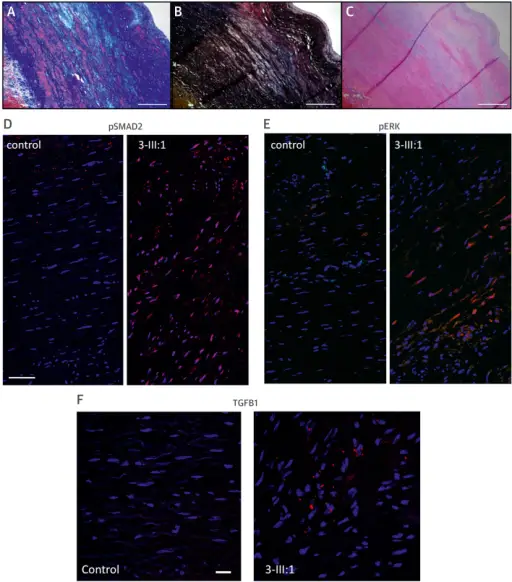
Cardiovascular Pathology and Immunohistochemical Analysis of TGFB Family Proteins in a Human Subject With TGFB3 Mutation (3-III:1; p.Asp263His)(A) Masson trichrome staining shows increased deposition of collagen (dark blue) and loss of smooth muscle fibers (red) in the media. (B) Elastin stain (Elastica van Gieson) shows loss of elastin fibers (black). (C) Hematoxylin-eosin staining shows deposition of proteoglycan (light blue) in the media. (A–C) Scale bar indicates 2 mm. (D–F) Cross sections of the media of the aortic wall of patient 3-III:1 and a matched control. Red staining corresponds to pSmad2 (D); pERK (E); and TGFB1 (F). Scale bars indicate 50 μm (D–E), 20 μm (F). Blue staining shows cell nuclei (DAPI), colocalization is purple. Red staining not colocalized with DAPI is nonspecific. Mutations in a TGF-β ligand, TGFB3, cause syndromic aortic aneurysms and dissections. Bertoli-Avella AM, Gillis E, Morisaki H, Verhagen JM, de Graaf BM, van de Beek G, Gallo E, Kruithof BP, Venselaar H, Myers LA, Laga S, Doyle AJ, Oswald G, van Cappellen GW, Yamanaka I, van der Helm RM, Beverloo B, de Klein A, Pardo L, Lammens M, Evers C, Devriendt K, Dumoulein M, Timmermans J, Bruggenwirth HT, Verheijen F, Rodrigus I, Baynam G, Kempers M, Saenen J, Van Craenenbroeck EM, Minatoya K, Matsukawa R, Tsukube T, Kubo N, Hofstra R, Goumans MJ, Bekkers JA, Roos-Hesselink JW, van de Laar IM, Dietz HC, Van Laer L, Morisaki T, Wessels MW, Loeys BL - Journal of the American College of Cardiology (2015). Not Altered. CC.
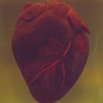
Cardiovascular pathology is usually caused by a buildup of fatty plaques in your arteries (atherosclerosis).
Examples of cardiovascular pathology include:
- Myocardial infarction
- Pulseless electrical activity
 What is Septic Shock?