Lentigo is a small sharply circumscribed pigmented macule surrounded by normal appearing skin.
What is the Pathology of Lentigo?
The pathology of lentigo is:
-Etiology: The cause of lentigo is dependent on the cause it can be solar lentigo, radiation lentigo and can be caused by genetic factors.
-Genes involved: None.
-Pathogenesis: The sequence of events that lead to lentigo is dependent on the cause in the solar lentigo. The UV light causes overproduction of melanin and the production of abnormal pigment retention.
-Morphology: The morphology associated with lentigo shows irregular or regular shaped macule that is darker in color than the normal skin color.
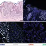
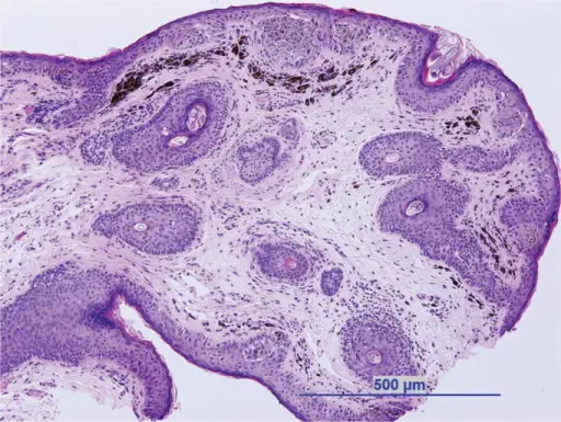
-Histology: The histology associated with lentigo shows increased basal melanin, increased number of melanocytes elongation of the rete ridges.
How does Lentigo Present?
Patients with lentigo are typically more common in males than in females and can be found in both children and adults. The symptoms, features, and clinical findings associated with lentigo include painless, redness at times.
How is Lentigo Diagnosed?
The lentigo is diagnosed physical exam and history taking examination with a wood lamp.
How is Lentigo Treated?
The lentigo is treated topical medication, cryosurgery, laser surgery.
What is the Prognosis of Lentigo?
The prognosis of lentigo is good.