Malignant fibrous histiocytoma is pleomorphic sarcoma composed of fibroblasts, myofibroblasts, and histiocyte-like cells.
What is the Pathology of Malignant Fibrous Histiocytoma?
The pathology of malignant fibrous histiocytoma is:
Etiology: The cause of malignant fibrous histiocytoma is not clear. Although these tumors may be linked to other medical conditions such as Paget disease, certain chemotherapy treatments, or past radiation treatments.
-Genes involved: G1/S checkpoint genes.
-Pathogenesis: The sequence of events that lead to malignant fibrous histiocytoma has not been clarified to date. However, it has been recognized as a complication of radiation, resulting from chronic postoperative repair, trauma, surgical incisions, or burn scars.
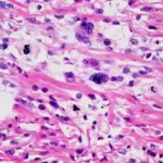
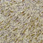
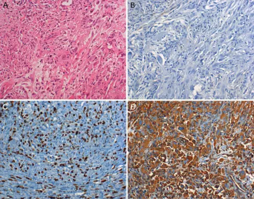
-Histology: The histology associated with malignant fibrous histiocytoma shows high cellularity, marked nuclear pleomorphism, usually accompanied by abundant mitotic activity and a spindle cell morphology.
How does Malignant Fibrous Histiocytoma Present?
Patients with malignant fibrous histiocytoma typically affect males slightly more than females present at an age of more than 50 years. The symptoms, features, and clinical findings associated with malignant fibrous histiocytoma include painless, enlarging nodules that can become painful if enlarging rapidly.
How is Malignant Fibrous Histiocytoma Diagnosed?
Malignant fibrous histiocytoma is diagnosed through MRI and biopsy.
How is Malignant Fibrous Histiocytoma Treated?
Malignant fibrous histiocytoma is treated with wide local excision.
What is the Prognosis of Malignant Fibrous Histiocytoma?
The prognosis of malignant fibrous histiocytoma is good since it is superficial, small in size, and low grade.