Nodular fasciitis is a benign, soft tissue tumor.
What is the Pathology of Nodular Fasciitis?
The pathology of nodular fasciitis is:
-Etiology: The cause of nodular fasciitis is unknown. However, it is thought to be the local reaction to traumatic injuries.
-Genes involved: USP-6 and MYH-9.
-Pathogenesis: The sequence of events that lead to nodular fasciitis involves the self-limiting growth of a clone of neoplastic cells that contain a fusion gene.
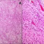
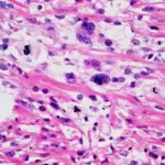
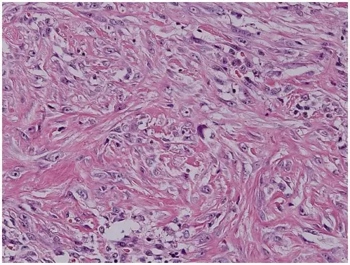
-Histology: The histology associated with nodular fasciitis shows spindle-shaped myofibroblastic cells.
How does Nodular Fasciitis Present?
Patients with nodular fasciitis typically affect both males and females present at an age range of 20-40 years. The symptoms, features, and clinical findings associated with nodular fasciitis show mild pain, discomfort, or soreness.
How is Nodular Fasciitis Diagnosed?
Nodular fasciitis is diagnosed through a sonogram, MRI, CT scan, and biopsy.
How is Nodular Fasciitis Treated?
Nodular fasciitis is treated by simple excision.
What is the Prognosis of Nodular Fasciitis?
The prognosis of nodular fasciitis is excellent.