Hemangiomas are usually benign vascular tumors derived from blood vessel cell types.
What is the Pathology of Hemangioma?
The pathology of hemangioma is:
-Etiology: The cause of hemangioma is not currently well-established.
-Genes involved: TEM8 (ANTXR1), VEGFR2, VEGFR3 (FLT4), and DUSP5.
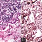
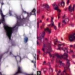
-Pathogenesis: The pathogenesis of hemangiomas is characterized by endothelial and pericytic hyperplasia, followed by a slower but steady involution phase that may last for years.
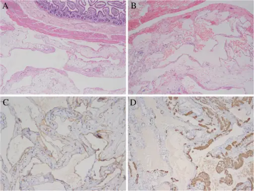
-Histology: The histology associated with hemangiomas shows hyperplastic endothelial cells, pericytes with and without lumens, and prominent basement membranes.
How does Hemangioma Present?
Patients with hemangioma typically affect females five times more than males during infancy. The symptoms, features, and clinical findings associated with hemangioma include flat and erythematous red patches.
How is Hemangioma Diagnosed?
Hemangioma is diagnosed clinically through biopsy and CT scan.
How is Hemangioma Treated?
Hemangioma is treated through beta-blockers and corticosteroids.
What is the Prognosis of Hemangioma?
The prognosis of hemangioma is very good.