The vascular wall response to injury is a condition of the blood vessels that is characterized by endothelial cell loss or dysfunction triggers the recruitment of smooth muscle cells, which proliferate; and an associated increase in the synthesis of matrix products resulting in intimal thickening.
What is the Vascular Wall Response to Injury?
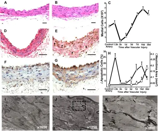
Early-onset apoptosis precedes vascular remodelling after arterial injury in rats(A and B) Haematoxylin and eosin stained cross sections from control (A) and injured (B) iliac arteries harvested at 90 min after surgery. (C) Temporal changes in the number of medial VSMC after vascular injury. (D and E) Detection of apoptotic VSMC in control (D) and injured (E) arteries at 90 min using a combination of In Situ Oligo Ligation (brown) to detect apoptotic nuclei and immunohistochemistry for SMA (red). Representative apoptotic VSMC are shown by arrows. (F and G) Representative microphotographs of control (F) and injured (G) arteries harvested at 90 min after surgery and stained for activated caspase 3. (H) Temporal quantification of apoptotic cells and neointima formation in the rat injured artery. Each point represents the mean ± S.E.M. (n=5-7). (I–K) Transmission electron microscopy of a normal (I) and injured artery (J). Apoptotic nuclei in the box are magnified in panel K. Scale bars=50 μm.Oxidative stress induces early-onset apoptosis of vascular smooth muscle cells and neointima formation in response to injury.
Gomez C, Martinez L, Mesa A, Duque JC, Escobar LA, Pham SM, Vazquez-Padron RI - Bioscience reports (2015). Not Altered. CC.
Post navigation
Previous Post
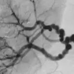 What is Fibromuscular Dysplasia?