Acute Leukemia
Acute Leukemia Pathology Video
Acute leukemia is the neoplastic proliferation of blast cells.
The abnormal blood cells in acute leukemia are immature blood cells called blasts.
Acute leukemia is defined as the accumulation of greater than 20% blasts in the bone marrow.
Acute leukemia typically presents as:
- Anemia
- Fatigue
- Thrombocytopenia
- Bleeding
- Neutropenia
The white blood cell (WBC) count is often high when blasts enter the bloodstream.
Blasts are large, immature cells that frequently have nucleoli that look punched out.
Based on the phenotype of the blasts, acute leukemia is further separated into two subdivisions:
- Acute lymphoblastic leukemia (ALL)
- Acute myelogenous leukemia (AML)
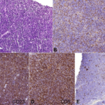
 T-lymphoblastic leukemia. Peripheral blood smear containing leukemic cells (Wright's stain, ×200) (A). Bone marrow aspirate smear showing leukemic cells that are small-to-medium sized, with a high nuclear-to-cytoplasmic ratio (Wright-Giemsa stain, ×200 (B) and ×1,000 (C)). Biopsy section showing hypercellularity with heavy infiltration of immature cells (H&E, ×100) (D). Simultaneous translocation of both TCR Loci (14q11) with rare partner loci (Xq22 and 12p13) in a case of T-lymphoblastic leukemia. Kang DH, Kim SH, Jun JW, Lee YW, Shin HB, Ahn JY, Hong DS, Lee YK, Jeon BR - Annals of laboratory medicine (2012). Not altered. CC.

Acute Lymphoblastic Leukemia (ALL)
Acute lymphoblastic leukemia (ALL) is a malignancy in the blood and bone marrow.
Acute lymphoblastic leukemia (ALL) is defined as neoplastic accumulation of lymphoblasts greater than 20%.
A DNA polymerase called terminal deoxynucleotidyl transferase (TdT) positive nuclear staining identifies lymphoblasts.
Myeloid blasts and mature lymphocytes do not contain terminal deoxynucleotidyl transferase (TdT).
Children are most frequently affected by acute lymphoblastic leukemia (ALL).
Patients with down syndrome are at increased risk of acute lymphoblastic leukemia (ALL).
Acute lymphoblastic leukemia (ALL) is linked to down syndrome, which typically manifests after the age of five.
Based on surface markers, acute lymphoblastic leukemia (ALL) is further classified into:
- T-cell lineage (T-ALL)
- B-cell lineage (B-ALL)
B cell acute lymphoblastic leukemia (B-ALL) is more common than T cell acute lymphoblastic leukemia (T-ALL).
B cell acute lymphoblastic leukemia (B-ALL)
B-ALL is characterized by lymphoblasts (TdT+) that also express CD10, CD19, and CD20.
B-ALL displays a fantastic response to chemotherapy.
B-ALL treatment requires prophylaxis to scrotum and CSF.
Cytogenetic abnormalities are useful to gauge the prognosis of B-ALL.
B-ALL with t(12;22) is seen in children more than adults, and has a good prognosis.
Philadelphia chromosome positive B-ALL (Ph+ B-ALL) is seen in adults more than children, and has a poor prognosis.
T cell acute lymphoblastic leukemia (T-ALL)
T-ALL is distinguished by lymphoblasts (TdT+) that also express CD2, CD3, CD5, CD7, and CD8.
Acute lymphoblastic lymphoma, so named as mediastinal (thymic) mass, typically manifests in teenagers because the malignant cells form a mass.
 Acute Lymphoblastic Leukemia. Acute lymphoblastic leukemia (ALL), peripheral blood of a child, Pappenheim stain, magnification x100. Christaras A. Not altered. CC BY 2.5


Acute Myeloid Leukemia (AML)
Acute myeloid leukemia (AML) is a malignancy of the blood and bone marrow.
Acute myeloid leukemia (AML) is a neoplastic accumulation of myeloblasts greater than 20%.
Myeloperoxidase positive (MPO) cytoplasmic staining is typically a feature of myeloblasts.
Crystal aggregates of myeloperoxidase positive (MPO) may be seen as Auer rods.
Auer rods are needle shaped large crystals which are cytoplasmic inclusion bodies, and are occasionally seen in myeloid blast cells during acute myeloid leukemia.
Acute myeloid leukemia (AML) typically strikes elderly people between the ages of 50 and 60 years old.
Acute myeloid leukemia (AML) is sub-classified based on surface markers, and cytogenetic abnormalities.
Acute myeloid leukemia (AML) can also develop from pre-existing dysplasia (myelodysplastic syndromes), particularly in cases when alkylating drugs or radiation therapy were previously used.
The typical findings of acute myeloid leukemia (AML) include:
- Cytopenias
- Hypercellular bone marrow
- Abnormal myeloid cell maturation
- Increased proportion of blast cells (greater than 20%)
Patients with acute myeloid leukemia (AML) are at risk of death due to infection or hemorrhage.
 Acute Myeloid Leukemia. Swollen gums due to infiltration by leukemia cells in a person with AML Herbert L. Fred, MD and Hendrik A. van Dijk - Not altered. CC BY-SA 3.0

Acute Promyelocytic Leukemia
Acute promyelocytic leukemia (APL) is a subtype of acute myeloid leukemia (AML) that involves translocation of the retinoic acid receptor (RAR) on chromosome 17 to chromosome 15.
When retinoic acid receptor (RAR) is disrupted, maturation is prevented and promyelocytes (blasts) accumulate.
Acute promyelocytic leukemia is treated with vitamin A derivative all-trans retinoic acid (ATRA) is used for treatment.
The vitamin A derivative all-trans-retinoic acid (ATRA), binds to the altered receptor and induces the blasts to mature.
 Acute Promyelocytic Leukemia. BONE MARROW: HYPERGRANULAR ACUTE åPROMYELOCYTIC LEUKEMIA (AML-M3) Postchemotherapy bone marrow smear from a patient with hypergranular acute promyelocytic leukemia. A single "faggot" cell with numerous Auer rods is present in a background of normal neutrophil precursors. (Wright-Giemsa stain) The Armed Forces Institute of Pathology (AFIP) - PEIR Digital Library (Pathology image database). Image# 404567. Not altered. Public Domain.

Acute Monocytic Leukemia
Acute monocytic leukemia is a subtype of acute myeloid leukemia (AML).
Acute monocytic leukemia is characterized by the growth of monoblasts and typically lacks MPO.
These blasts characteristically infiltrate gums.
Acute Megakaryoblastic Leukemia
Acute megakaryoblastic leukemia is a subtype of acute myeloid leukemia (AML).
Acute megakaryoblastic leukemia is characterized by proliferation of megakaryoblasts that express platelet-specific surface glycoproteins.
Acute megakaryoblastic leukemia lacks myeloperoxidase positive (MPO).
Acute megakaryoblastic leukemia is connected to Down syndrome, which often manifests before the age of five years old.