Amyloidosis
Amyloid refers to various fibrous, extracellular, proteinaceous deposits.
Amyloidosis is the abnormal buildup of amyloid in various tissues of the body.
Types of amyloidosis include:
- AA amyloidosis
- Hereditary amyloidosis (familial amyloidosis)
- Wild-type amyloidosis
- Localized amyloidosis
- AL amyloid (immunoglobulin light chain amyloidosis)
The beta-pleated sheet configuration of amyloid is common among amyloidosis.
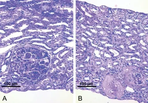
The classic stain for amyloid is Congo red which under polarized light has a characteristic apple green birefringence when viewed microscopically.
The deposition of amyloid may occur localized or systemically.

Localized Amyloidosis
Localized amyloidosis is defined as amyloid deposition in a single organ.
Examples of localized amyloidosis include:
- Senile cardiac amyloidosis (non-mutated serum transthyretin)
- Familial amyloid cardiomyopathy (mutated serum transthyretin)
- Type 2 diabetes mellitus (amylin from excess insulin)
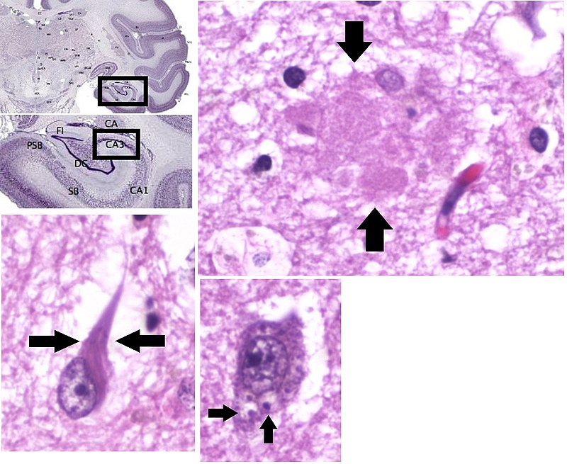
- Alzheimer disease (AB amyloid)
- Dialysis associated amyloidosis (B2-microglobulin)
- Medullary carcinoma of the thyroid (calcitonin)
Senile cardiac amyloidosis is due to buildup of non-mutated serum transthyretin in the heart that may lead too restrictive cardiomyopathy.
Familial amyloid cardiomyopathy is due to mutated serum transthyretin that deposits in the heart causing restrictive cardiomyopathy.
Type 2 diabetes mellitus may cause amyloidosis by amylin being deposited in the pancreas islets.
Alzheimer disease is associated with AB amyloid depositing and forming amyloid plaques in the brain.
Dialysis associated amyloidosis is associated with B2-microgobulin depositing in joints.
Medullary carcinoma of the thyroid results in calcitonin deposition in the tumor in an amyloid like fashion.
Systemic Amyloidosis
Systemic amyloidosis results in systemic deposition of amyloid.
Systemic amyloidosis may be due to primary amyloidosis or secondary amyloidosis.
Primary amyloidosis: AL amyloid, which originates from the immunoglobulin light chains.
Secondary amyloidosis: AA amyloid, which is derived from SAA, an acute phase reactant that is increased in certain conditions.
Conditions that result in SAA include:
- Familial Mediterranean fever (due to dysfunctioning neutrophils attacking serosal surfaces)
- Malignancy
- Chronic inflammation
Primary amyloidosis is associated with multiple myeloma and other plasma cell dyscrasia such as plasmacytomas and monoclonal gammopathy of undetermined significance (MGUS).
Secondary amyloidosis is systemic deposition of a amyloid which is originating from the serum amyloid associated protein.
Serum amyloid associated protein is an acute phase reactant associated with chronic inflammation and cancer.
Clinical conditions associated with systemic amyloidosis include:
- Nephrotic syndrome (most commonly)
- Restrictive cardiomyopathy
- Hepatosplenomegaly
- Macroglossia (tongue enlargement)
- Thickened bowel wall
Nephrotic syndrome is a kidney disorder that causes proteinuria.
The diagnosis of amyloidosis is made by biopsy.
Amyloid cannot be removed.
Organs damaged by amyloidosis must be transplanted.