Anterior Pituitary Gland Pathology Video
Pituitary Adenoma
Pituitary adenoma is a benign tumor of anterior pituitary cells.
Pituitary adenomas may be functional meaning it is hormone-producing.
Pituitary adenomas may be nonfunctional meaning it is silent.
Nonfunctional tumors often present with mass effect.
Bitemporal hemianopia may occur due to compression of the optic chiasm.
Compressed normal pituitary tissue causes hypopituitarism.
Based on the type of hormone produced, functional tumors have certain characteristics.
Prolactinoma
Prolactinoma presents as galactorrhea and amenorrhea in females or as decreased libido and headache in males.
Among the majority of pituitary tumors, about 30% of them are prolactin-secreting pituitary adenomas which is considered as the most common type.
The treatment used for this condition are dopamine agonists such as bromocriptine or cabergoline to suppress prolactin production, which shrinks the tumor, or surgery when lesions are larger.
Growth hormone (GH) cell adenoma
Growth hormone cell adenoma causes gigantism in children characterized by increased linear bone growth (epiphyses are not fused).
They have high levels of growth hormones in the body, which cause them to become unusually tall.
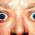
Growth hormone cell adenoma causes acromegaly in adults characterized with enlarged bones of hands, feet, and jaw.
This happens when the body produces excessive growth hormone in adults.
The growth of visceral organs may cause the progression of organ dysfunction, cardiac failure.
Secondary diabetes mellitus is often present (growth hormone (GH) induces liver gluconeogenesis).
It is diagnosed by elevated growth hormone (GH) and insulin growth factor-1 (IGF-1) levels along with lack of growth hormone suppression by oral glucose.
Octreotide, a somatostatin analog that suppresses growth hormone (GH) release, is the drug of choice for treatment of growth hormone cell adenoma.
Growth hormone receptor antagonists and surgery may be necessary.
Adrenocorticotropic hormone releasing tumors
Adrenocorticotropic hormone (ACTH) releasing tumors are a type of adenoma that secrete ACTH leading to Cushing syndrome.
Cushing syndrome is a condition that develops when the body consistently produces excessive amounts of the hormone cortisol.
Thyroid-stimulating hormone (TSH) cells, luteinizing hormone (LH)-producing, and follicle-stimulating hormone (FSH)-producing adenomas occur, but are rare.
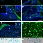
 Pituitary Adenoma. True null cell adenomas are typically composed of uniform, mildly atypical cells with chromophobic cytoplasm. This case has papillary architecture similar to gonadotroph adenomas.[53] Juliana Drummond, Federico Roncaroli, Ashley B. Grossman, and Ma´ rta Korbonits. not altered. CC BY 4.0


Hypopituitarism
Hypopituitarism is a condition wherein the anterior pituitary gland is not producing enough hormones.
Symptoms of hypopituitarism arise when more than 75% of the pituitary parenchyma is lost.
Causes of hypopituitarism include:
- Pituitary adenomas in adults or craniopharyngioma in children – this is caused by the mass effect or pituitary apoplexy which is the bleeding into an adenoma.
- Sheehan’s syndrome commonly known as the postpartum pituitary necrosis; this condition describes the infarction of anterior pituitary gland cells.
Although the size of the gland increases during pregnancy, the blood supply does not greatly increase.
Parturition related blood loss causes an infarction to occur.
Symptoms of hypopituitarism include:
- Poor lactation
- Pubic hair loss
- Fatigue
Empty sella syndrome is a rare disorder when the pituitary gland flattens or decreases as a result of problems with the sella turcica’s contents.
Herniation of the arachnoid and cerebral spinal fluid (CSF), otherwise known as cerebrospinal fluid, into the sella turcica compresses and destroys the pituitary gland.
The pituitary gland is “absent” which indicates that the sella turcica is empty on imaging.
 Hypopituitarism. Pituitary Gland Henry Vandyke, Henry Not altered. Public Domain