Central Nervous System Developmental Anomalies
Central Nervous System Developmental Pathology Video
Neural Tube Defects
The incomplete closure of the neural tube causes neural tube defects (NTDs).
Early in gestation, the neural plate invaginates to create the neural tube, which follows the embryo’s cranial-caudal axis.
The peripheral nervous system is formed by the neural crest.
The ventricles and spinal cord canal are formed by the hollow lumen of the neural tube and its wall.
Neural tube defects (NTDs) can be caused by low folate levels prior to conception.
Types of neural tube defects (NTDs) include:
- Anencephaly
- Spina bifida
- Meningocele
- Meningomyelocele
Anencephaly
An absence of the skull and brain caused by a disruption of the neural tube’s cranial end is known as anencephaly.
As a result, the fetus develops a “frog-like” look and the mother develops polyhydramnios due to the fetus’s decreased ability to consume amniotic fluid.
Spina bifida
Spina bifida is a vertebral defect caused by the posterior vertebral arch’s inability to close (disruption of the caudal end of the neural tube).
Spina bifida occulta presents as a dimple or patch of hair overlying the vertebral defect.
Cystic extrusion of the underlying tissue through the spinal defect is how spina bifida manifests.
Meningocele
Meningocele is a congenital abnormality that is characterized by a sac that protrudes from the spinal column of the meninges.
Meningomyelocele
Meningomyelocele is the protrusion of meninges and spinal cord.
During prenatal care, high alpha-fetoprotein (AFP) levels in the amniotic fluid and the mother’s blood can be used to identify neural tube defects.
 Neural Tube Defects. An illustration of an infant with Spina Bifida Centers for Disease Control and Prevention - Centers for Disease Control and Prevention. Not altered. Public Domain

Cerebral Aqueduct Stenosis
In order to understand cerebral aqueduct stenosis, you must first understand how cerebrospinal fluid (CSF) is produced and how it flows:
- Cerebrospinal fluid (CSF) is produced by the choroid plexus lining the ventricles
- Flows from the lateral ventricles into the 3rd ventricle via the interventricular foramen of Monro
- Flows from the third ventricle into the fourth ventricle via the cerebral aqueduct
- Flows from the fourth ventricle into the subarachnoid space via the foramina of Magendie and Luschka
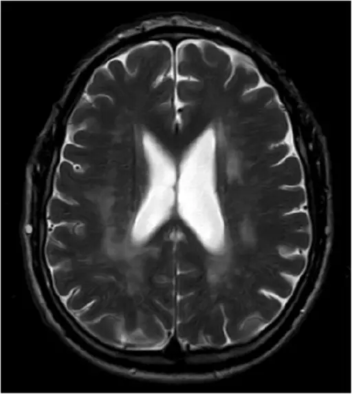
In cerebral aqueduct stenosis, cerebrospinal fluid (CSF) is drained from the third ventricle into the fourth ventricle through a conduit that has a congenital stenosis, thus blocking the flow of CSF.
Cerebral aqueduct stenosis leads to accumulation of cerebrospinal fluid (CSF) in the ventricular space.
Cerebral aqueduct stenosis often causes hydrocephalus in newborns.
Cerebral aqueduct stenosis presents with enlarging head circumference due to dilation of the ventricles (cranial suture lines are not fused).
 Cerebral Aqueduct Stenosis. The enlarged skull of a person with hydrocephalus, which is a symptom of the excess CSF in the ventricular system. This may be caused by aqueductal stenosis, and in some cases, it is thought that hydrocephalus will cause aqueductal stenosis. Otis Historical Archives of “National Museum of Health & Medicine” (OTIS Archive 1) - Not altered. CC BY 2.0

Dandy Walker Malformation
A congenital brain deformity called Dandy-Walker malformation affects the cerebellum, which controls movement, and the fluid-filled areas around it.
Dandy-Walker malformation is a congenital failure of the cerebellar vermis to develop.
A significantly dilated fourth ventricle (posterior fossa) with an absent cerebellum and hydrocephalus are the hallmarks of this Dandy-Walker malformation.
 Dandy Walker Malformation. Diagram of the cerebellum, fourth ventricle and pons. The white arrow shows the foramen of Magendie (medial aperture) connecting the fourth ventricle to the cisterna magna (3). This usually remains open in DWM.[6] lyhana8 - Not altered. Public Domain


Arnold Chiari Malformation
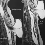
Arnold-Chiari malformation is a condition in which the spinal canal is invaded by the lower section of the brain.
Arnold-Chiari malformation is a foramen magnum-based congenital expansion of the cerebellar tonsils.
Hydrocephalus may develop in Arnold-Chiari malformation as a result of cerebrospinal fluid (CSF) flow obstruction.
Arnold-Chiari malformation may present together with meningomyelocele or syringomyelia.
 Arnold Chiari Malformation. Syringomyelia associated with Chiari malformation © Nevit Dilmen Dichromatic, False-color MRI. Cervical spine, T1 Red, T2, Green& Blue Permission details You may select the license of your choice. Not altered. CC BY-SA 3.0