Chronic Leukemia
Chronic Leukemia Pathology Video
Chronic leukemia involves leukemia of mature blood cells.
Chronic leukemia is typically insidious in onset and visible in elderly adults.
Examples of chronic leukemias include:
- Chronic lymphocytic leukemia/small lymphocytic lymphoma (CLL/SLL)
- Hairy cell leukemia (HCL)
- Adult T-cell leukemia/lymphoma
- Mycosis fungoides (MF)
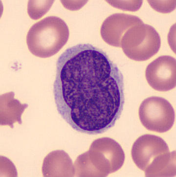
 Chronic Myeloid Leukemia. Peripheral blood (MGG stain): marked leukocytosis with granulocyte left shift Paulo Henrique Orlandi Mourao - Not altered. CC BY-SA 3.0


Chronic Lymphocytic Leukemia (CLL)
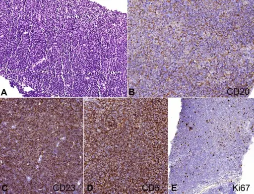
Chronic lymphocytic leukemia/small lymphocytic lymphoma (CLL/SLL) is characterized by B cells that co-express CD5 and CD20.
Chronic lymphocytic leukemia/small lymphocytic lymphoma (CLL/SLL) is the most prevalent type of chronic leukemia.
Chronic lymphocytic leukemia/small lymphocytic lymphoma (CLL/SLL) classically shows smudge cells and lymphocytes on blood smear.
Small lymphocytic lymphoma is characterized by lymph node involvement, which results in widespread lymphadenopathy.
Complications of chronic lymphocytic leukemia/small lymphocytic lymphoma (CLL/SLL) include:
- Autoimmune hemolytic anemia
- Hypogammaglobulinemia
- Risk of infections
- Transformation to diffuse large B-cell lymphoma (Richter transformation)
The leading cause of death in chronic lymphocytic leukemia/small lymphocytic lymphoma (CLL/SLL) is hypogammaglobulinemia compounded by infection.
 Chronic Lymphocytic Leukemia. Smudge cells in peripheral blood Dr Graham Beards - Not altered. CC BY-SA 3.
Hairy Cell Leukemia (HCL)
Hairy cell leukemia (HCL) is characterized by cytoplasmic processes that radiate from the malignant cells.
Hairy cell leukemia (HCL) is positive for tartrate-resistant acid phosphatase (TRAP) stain.
Symptoms of hairy cell leukemia (HCL) includes:
- Splenomegaly (caused by an increase of hairy cells in the red pulp)
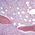
- Fibrotic “dry tap” bone marrow aspiration
Lymphadenopathy is normally absent in hairy cell leukemia (HCL).
Hairy cell leukemia (HCL) is treated by adenosine deaminase inhibitor 2-chlorodeoxyadenosin (2-CdA).
The adenosine deaminase inhibitor 2-chlorodeoxyadenosin (2-CdA) causes the neoplastic B cells to accumulate adenosine to lethal amounts.
2-chlorodeoxyadenosin (2-CdA) is also known as Cladribine (aka Leustatin).
If treated hairy cell leukemia (HCL) has an excellent prognosis.
 Hairy Cell Leukemia. Atypical hairy cell lymphocytes (arrows) seen in peripheral blood. Hairy cell leukemia - immunotargets and therapies. Basheer F, Bloxham DM, Scott MA, Follows GA - ImmunoTargets and therapy (2014). Not altered. CC.


Adult T-Cell Leukemia/Lymphoma (ATLL)
Adult T-cell leukemia/lymphoma is due to neoplastic proliferation of mature T cells.
Adult T-cell leukemia/lymphoma is linked to HTLV-1.
Adult T-cell leukemia/lymphoma is most prevalent in Japan and the Caribbean.
Symptoms of Adult T-cell leukemia/lymphoma include:
- Rashes
- Generalized lymphadenopathy
- Hepatomegaly
- Splenomegaly
- Lytic (punched-out) bone lesions
- Hypercalcemia
 T-lymphoblastic leukemia. Peripheral blood smear containing leukemic cells (Wright's stain, ×200) (A). Bone marrow aspirate smear showing leukemic cells that are small-to-medium sized, with a high nuclear-to-cytoplasmic ratio (Wright-Giemsa stain, ×200 (B) and ×1,000 (C)). Biopsy section showing hypercellularity with heavy infiltration of immature cells (H&E, ×100) (D). Simultaneous translocation of both TCR Loci (14q11) with rare partner loci (Xq22 and 12p13) in a case of T-lymphoblastic leukemia. Kang DH, Kim SH, Jun JW, Lee YW, Shin HB, Ahn JY, Hong DS, Lee YK, Jeon BR - Annals of laboratory medicine (2012). Not altered. CC.


Mycosis Fungoides (MF)
Mycosis fungoides (MF) is due to the neoplastic proliferation of mature CD4+ T cells that infiltrate the skin.
Symptoms of mycosis fungoides (MF) includes:
- Skin rashes (nodules and plaques)
- Fatigue
- Hepatomegaly
Histology of mycosis fungoides (MF) is characterized by malignant infiltration of neoplastic-T-cells in the epidermis that are known as Pautrier microabscesses.
 Mycosis Fungoides. Histopathology of Pautrier microabscesses in cutaneous T cell lymphoma. Nooshin Bagherani, Bruce R. Smoller - Not altered. CC BY 4.0


Sézary Syndrome
Sézary syndrome is a type of mycosis fungoides (MF) in which the malignant cells also circulate in the blood stream.
On a blood smear, Sézary syndrome characteristically shows lymphocytes with cerebriform nuclei (Sézary cells).