Endocarditis
Endocarditis is inflammation of the endocardium.
Endocarditis is typically brought on by a bacterial infection.
Streptococcus viridans is the most frequent cause of endocarditis.
Streptococcus viridans is a low-virulence bacteria that spreads to damaged valves.
Damaged cardiac valves are commonly due to chronic rheumatic heart disease and mitral valve prolapse.
Rheumatic heart disease
Leads to small vegetations that don’t destroy the valve (subacute endocarditis).
Thrombotic vegetations form on damaged endocardial surfaces (platelets and fibrin).
Temporary bacteremia causes microorganisms to become trapped in vegetation.
Prophylactic antibiotics reduce the risk of endocarditis.
Common associations of endocarditis include:
- In IV drug users, Staphylococcus aureus is the most frequent cause
- Prosthetic valve endocarditis is linked to Staphylococcus epidermidis
- Streptococcus bovis in patients with underlying colorectal carcinoma
High-virulence organism may infects normal valves, most frequently the tricuspid valve, leading to large vegetations that destroy the valve (acute endocarditis).
HACEK organisms are linked to endocarditis with negative blood cultures.
HACEK organisms include:
- Haemophilus
- Actinobacillus
- Cardiobacterium
- Eikenella
- Kingella
Clinical features of bacterial endocarditis are:
- Fever (because of bacteremia)
- Murmur (because of vegetations on the heart valve)
- Janeway lesions (erythematous non tender lesions on the palms and soles)
- Osler nodes (tender lesions on fingers or toes), and splinter hemorrhages in nail bed (because of embolization of septic vegetation)
- Anemia of chronic disease (due to hepcidin binding up iron in the face of chronic inflammation)
Laboratory findings of bacterial endocarditis include:
- Positive blood cultures
- Anemia of chronic disease
Transesophageal echocardiography is helpful for evaluating the cardiac valve lesions.
Sterile vegetations that develop in conjunction with a hypercoagulable condition or an underlying adenocarcinoma are the cause of nonbacterial thrombotic endocarditis.
Mitral regurgitation is brought on by vegetation that grows along the lines of closure on the mitral valve.
Libman-Sacks endocarditis is a nonbacterial thrombotic endocarditis that is associated with systemic lupus erythematous (SLE).
The mitral valve has vegetation on its surface and underneath, which causes mitral regurgitation.
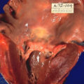
 Endocarditis Pathology. ID#: 851 Description: Gross pathology of subacute bacterial endocarditis involving mitral valve. Left ventricle of heart has been opened to show mitral valve fibrin vegetations due to infection with Haemophilus parainfluenzae. Autopsy. Content Providers(s): CDC/Dr. Edwin P. Ewing, Jr. Creation Date: 1972 Copyright Restrictions: None - This image is in the public domain and thus free of any copyright restrictions. As a matter of courtesy we request that the content provider be credited and notified in any public or private usage of this image. http://phil.cdc.gov/PHIL_Images/02122002/00007/PHIL_851_lores.jpg. Not altered. Public Domain