Gestational pathology is pathology of the placenta or embryo.
Gestational pathology includes:
- Ectopic pregnancy
- Spontaneous abortion
- Placenta previa
- Placental abruption
- Placenta accreta
- Preeclampsia
- Eclampsia
- Hemolysis, elevated liver enzymes and low platelets (HELLP) syndrome
- Sudden infant death syndrome (SIDS)
- Hydatidiform mole
- Choriocarcinoma
Ectopic Pregnancy
Ectopic pregnancy happens when fertilized ovaries are being implanted anywhere other than the uterine wall.
Ectopic pregnancy most frequently occurs in the fallopian tube which transmits eggs from the ovaries to the uterus is the site of an ectopic pregnancy.
Key risk factors for ectopic pregnancy include:
- Scarring the secondary to pelvic inflammatory disease
- Endometriosis
A few weeks after a missed period, women who have an ectopic pregnancy may experience irregular bleeding and lower abdominal pain or pelvic pain.
Ectopic pregnancy is considered a surgical emergency.
Major complications of ectopic pregnancy include:
- Bleeding into the fallopian tube (hematosalpinx)
- Rupture
 Ectopic Pregnancy. Laparoscopic view, looking from superiorly to inferiorly in the peritoneal cavity which has been pumped up with carbon dioxide gas to visualize the uterus (marked by blue arrows). On the left Fallopian tube there is an ectopic pregnancy and hematosalpinx (marked by red arrows). The right tube is normal. Mikael Häggström. Not altered. CC BY-SA 3.0

Spontaneous Abortion
Spontaneous abortion is fetal miscarriage that occurs before 20 weeks of pregnancy.
Spontaneous abortion typically occurs in the first trimester.
Spontaneous abortion is common and happens up to 1/4 of recognizable pregnancies.
Symptoms of spontaneous abortion include:
- Vaginal bleeding
- Cramp-like pain
- Passage of fetal tissues
Causes of spontaneous abortion include:
- Chromosomal anomalies
- Hypercoagulable states
- Antiphospholipid syndrome
- Teratogens
Effects of teratogens depend on the timeframe of exposure:
- First two weeks of gestation – spontaneous abortion
- Weeks 3-8 – risk of organ malformation
- Months 3-9 – risk of organ hypoplasia
 Spontaneous Abortion. Transvaginal ultrasonography after an episode of heavy bleeding in an intrauterine pregnancy that had been confirmed by previous ultrasonography. There is some widening between the uterine walls, but no sign of any gestational sac, thus, in this case, being diagnostic of a complete miscarriage. Mikael Häggström. Not altered. CC0

Placenta Previa
Placenta previa is a pregnancy problem that occurs when the placenta totally or partially blocks the uterine entrance (cervix).
Placenta previa usually presents as third-trimester bleeding.
Placenta previa often requires cesarean section, which is a procedure of delivering a baby through surgical incisions made in the abdomen and uterus.
Placental Abruption
Placental abruption is when the placenta is separated from the decidua before the fetus is delivered.
Placental abruption is a common cause of stillbirth.
The latter trimester of pregnancy, particularly the final few weeks before delivery, is when placental abruption is most prone to happen.
Symptoms of placenta abruption include:
- Uterine soreness
- Abdominal pain
- Vaginal bleeding
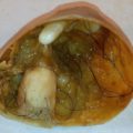
 Placental Abruption. Gross pathology of a uterus which has been opened to show a placental abruption, with a hematoma separating the placenta from the uterus. Mikael Häggström, M.D. Not altered. CC0
Placenta Accreta
Placenta accreta is a serious pregnancy condition wherein there is an improper placenta-myometrium implantation with minimal to no intervening decidua.
Placenta accreta is often recognized during labor with difficult delivery of the placenta and postpartum bleeding.
C-section and/or hysterectomy may be needed to treat placenta accreta.
 Placenta Accreta. A diagram illustrating the different types of placenta accreta. TheNewMessiah at English Wikipedia - Transferred from en.wikipedia to Commons. not altered Public Domain
Preeclampsia
Preeclampsia is edema, proteinuria, and hypertension are conditions brought on by pregnancy often appear in the third trimester and affect about 5% of pregnancies.
In preeclampsia the hypertension may be severe, leading to headaches and visual abnormalities.
Preeclampsia is due to an abnormality of the maternal-fetal vascular interface in the placenta.
Preeclampsia resolves with delivery.
 Preeclampsia. High magnification micrograph of hypertrophic decidual vasculopathy, as seen in pregnancy-induced hypertension. H&E stain. Related images Intermed. mag. Low mag. Nephron. Not altered. CC BY-SA 3.0


Eclampsia
Eclampsia is preeclampsia with seizures.
 Histological changes in placenta in cases of preeclampsia and eclampsia. Study of structural changes in placenta in pregnancy-induced hypertension. Salmani D, Purushothaman S, Somashekara SC, Gnanagurudasan E, Sumangaladevi K, Harikishan R, Venkateshwarareddy M - Journal of natural science, biology, and medicine (2014). Not Altered. CC.

HELLP Syndrome
Hemolysis, elevated liver enzymes and low platelets syndrome (HELLP syndrome) is preeclampsia in combination with liver-related thrombotic microangiopathy.
Both eclampsia and HELLP syndrome usually warrant immediate delivery.
Sudden Infant Death Syndrome (SIDS)
Sudden infant death syndrome (SIDS) occurs when infants ages one month old to one-year-old who suddenly died without apparent cause.
Sudden infant death syndrome is usually associated with issues with a baby’s inability to wake up and expire during sleep.
Risk factors for sudden infant death syndrome include:
- Exposure to cigarette smoke
- Prematurity
- Sleeping on stomach
 Sudden Infant Death Syndrome.SIDS rate from 1988 to 2006 This graph was obtained from here, the National Institute of Child Health and Human Development website. It is a product of a United States agency, the Department of Health and Human services, and is thus in the public domain and falls under no copyright. SIDS rates and back sleeping (1988-2006). Not altered Public Domain

")
Hydatidiform Mole
A hydatidiform mole is the condition that is marked by enlarged, swollen, and edematous villi and trophoblast growth.
A hydatidiform mole is known as molar pregnancy.
A hydatidiform mole causes the uterus to expand as though a normal pregnancy is present, but it is significantly larger and higher than predicted for the gestational age.
Hydatidiform moles are detected during prenatal care with routine ultrasonography in the early first trimester, which show absence of fetal heart sounds and the ultrasound classically shows a ‘snowstorm’ appearance.
A hydatidiform mole may be recognized on physical exam as a grape-like lumps passing through the vaginal canal.
Hydatidiform moles are classified as:
- Complete hydatidiform mole (Diploid, 45 XX)
- Partial hydatidiform mole (Triploid, 69 XXY)
Treatment of hydatid moles includes dilatation and curettage (D&C).
To guarantee adequate mole removal and to check for the emergence of choriocarcinoma, follow-up monitoring is crucial.
A hydatidiform mole, a normal pregnancy, or a spontaneous germ cell tumor can all result in choriocarcinoma as a gestational complication.
 Hydatidiform mole. Histopathogic image of hydatidiform mole (complete type). H & E stain.KGH (talk | contribs Not altered. CC BY-SA 3.0
Choriocarcinoma
Choriocarcinoma may also arise as a spontaneous germ cell tumor.
Choriocarcinoma is treated with chemotherapy.
Chemotherapy is more effective in treating choriocarcinomas that develop along the gestational pathway opposed to choriocarcinomas that develop via the germ cell pathway.
 Hematoxylin and eosin pathologic stain of epidural tumor. (a) Low-power view showing diffuse hemorrhages common in choriocarcinoma. (b) High-power view highlighting syncytiotrophoblasts and large, pleomorphic malignant single cells.Metastatic choriocarcinoma to the lumbar spine: Case report and review of literature. Skoch J, Kobylanski K, Rice JM, Baaj AA - Surgical neurology international (2014). Not Altered. CC