Ependymal cells react to injury by migrating to the site of injury and proliferating into neurons and glial cells in the adult spinal cord.
How do Ependymal Cells React to Injury?
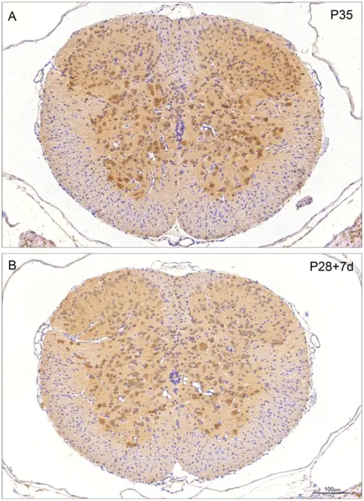
Immunocytochemical staining for ubiquitin in control (P35) and transected (P28+7 d) spinal cord of Monodelphis domestica. A: P35 control. Note ubiquitin staining similar to P29 (Figure 6A). Neuronal nuclei are as strongly stained as cytoplasm apart from Waldeyer cells at the tip of dorsal horn, which have stronger cytoplasmic staining. Some glial cells in white matter are positively stained. B: P28+7 d post injury. Note pattern of immunostaining is similar to that in age-equivalent control (P35, A) although less intense. Endothelial cells and neuroependymal cells lining the central spinal cord are negative. A and B, same magnification. Bar in B: 100 µm. Expression and cellular distribution of ubiquitin in response to injury in the developing spinal cord of Monodelphis domestica.
Noor NM, Møllgård K, Wheaton BJ, Steer DL, Truettner JS, Dziegielewska KM, Dietrich WD, Smith AI, Saunders NR - PloS one (2013). Not Altered. CC.
Post navigation
Previous Post
 How do Oligodendrocytes React to Injury?
Next Post
What is Cerebral Edema?