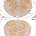
Oligodendrocytes react to injury with the hypertrophy and creating an inhibitory environment that prevents regeneration of axons.
How do Oligodendrocytes React to Injury?
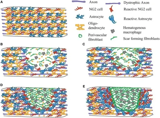
Scar formation after SCI. Diagram depicting the events of scar formation after contusive SCI in mice. Astrocytes (blue), NG2 cells (red), and myelinating oligodendrocytes (yellow) in the uninjured spinal cord white matter (A). Early after SCI, cell death occurs within the lesion site and axons are damaged. Microglia (not shown) and astrocytes respond by secreting cytokines and chemokines. NG2 cells react and proliferate around the lesion site. Macrophages (gray) begin to infiltrate the lesion core and perivascular fibroblasts (green) begin to delaminate from blood vessels (B). Inflammation causes secondary death of oligodendrocytes and neurons leading to accumulation of myelin debris in the injury site (C). Macrophage and fibroblast density peaks at 7 days after SCI (D). By 2 weeks after SCI, the scar has matured. There are tight borders between the fibrotic scar (consisting of fibroblasts and macrophages) and the glial scar (consisting of astrocytes, NG2 cells, and microglia) (E). The relative number of cells may not accurately reflect actual in vivo pathology. Understanding the NG2 Glial Scar after Spinal Cord Injury
Frontiers in Neurology. Not Altered. CC.
Post navigation
Previous Post
 How do Microglia React to Injury?