Langerhans Cell Histiocytosis
Langerhans Cell Histiocytosis Pathology Video
The skin is the primary location of Langerhans cells, which are specialized dendritic cells.
Langerhans cells are derived from bone marrow monocytes.
Langerhans cells present antigen to naive T cells.
A neoplastic growth of Langerhans cells is known as Langerhans cell histiocytosis (LCH).
Langerhans cell histiocytosis (LCH) takes place when the body produces an excessive number of immature Langerhans cells making it a cancer-like condition.
Tennis racket shaped structures known as Birbeck granules, are found in the Langerhans cell histiocytosis (LCH), and can be seen on electron microscopy.
Langerhans cell histiocytosis cells are CD1a+ by immunohistochemistry.
 Langerhans Cell Histiocytosis. Hand–Schüller–Christian disease. Not altered. CC BY-SA 3.0
Letterer-Siwe Disease
Letterer-Siwe disease is one of the variations of Langerhans cell histiocytosis (LCH), which is regarded as an uncommon condition, due to the malignant proliferation of Langerhans cells.
The disease is characterized by onset in infancy (less than 2-years-old) that shows skin rashes and cystic skeletal defects.
Multiple organs may be involved in Letterer-Siwe disease.
Letterer-Siwe disease is rapidly fatal.
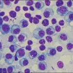
 Langerhans Cell Histiocytosis. Very high magnification micrograph of Langerhans cell histiocytosis, previously known as histiocytosis X, Hand-Schüller-Christian disease, and Abt-Letterer-Siwe disease. H&E stain. It is characterized by Langerhans type histiocytes which have a reniform (or kidney-shaped) nucleus and stain with S100 and CD1a. Related images Very low mag. Low mag. Intermed. mag. High mag. Nephron. Not altered CC BY-SA 3.0
Eosinophilic Granuloma
Eosinophilic granuloma is a lesion that typically affects the bones.
Eosinophilic granulomas are caused by an excess and overgrowth of Langerhans cells.
Pathologic fracture in an adolescent is the most common presentation of eosinophilic granulomas.
A biopsy and histologic assessment of eosinophilic granulomas reveals a mixture of inflammatory cells, including many eosinophils, along with Langerhans cells.
 Churg-Strauss Syndrome. High magnification micrograph of eosinophilic vasculitis consistent with Churg-Strauss syndrome, abbreviated CSS. H&E stain. CSS is characterized by: Granulomas. Asthma. Fever. Eosinophilia. Related images High mag. Very high mag. Nephron - Not altered. CC BY-SA 3.


Hand-Schüller-Christian Disease
Hand-Schüller-Christian disease is also known as chronic multifocal Langerhans cell histiocytosis.
The malignant growth of Langerhans cells causes Hand-Schüller-Christian disease.
Hand-Schüller-Christian disease is a condition in which the patient’s tissues or organs are attacked by histiocytes as they begin to multiply.
Classic presentation of Hand-Schüller-Christian disease involves:
- Scalp rash
- Lytic skull defects
- Diabetes insipidus
- Exophthalmos
 Hand-Schuller-Christian Disease. Not altered. CC BY-SA 3.0