Meningitis
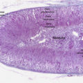
Meningitis is the technical term for inflammation of the leptomeninges.
The leptomeninges are made of three layers:
- Dura mater
- Arachnoid
- Pia mater
The meninges are located between the brain and the skull.
Leptomeninges is the collective word for pia and arachnoid.
Meningitis is typically caused by an infectious disease such as:
- Listeria monocytogenes, E coli, and Group B streptococci (neonates)
- Streptococcus pneumoniae (adults)
- Neisseria meningitidis (children and teenagers)
- Hemophilus influenza (unvaccinated infants)
- Coxsackievirus (children; fecal-oral transmission)
- Fungi (immunocompromised individuals)
Symptoms of meningitis include:
- Headache
- Nuchal rigidity
- Fever
- Photophobia
- Vomiting
- Altered mental state
Lumbar puncture is the diagnostic procedure for sampling of cerebrospinal fluid (CSF) to assess meningitis.
Lumbar puncture is performed by inserting a needle between L4 and L5 (level of the iliac crest).
Note that the spinal cord ends at L2, but subarachnoid space and cauda equina continue.
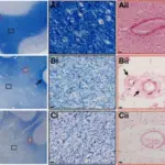
Cerebrospinal fluid (CSF) findings in meningitis depend on the cause.
Bacterial Meningitis
- Increased neutrophils
- Decreased glucose
The causative organism may be identified by gram stain, polymerase chain reaction (PCR), Matrix Assisted Laser Desorption/Ionization – Time of Flight (MALDI-TOF), and culture.
The majority of the time, bacterial meningitis has complications.
Viral Meningitis
- Increased lymphocytes
- Normal glucose
Fungal Meningitis
- Increased lymphocytes
- Increased glucose
Potential complications of meningitis include:
- Death due to herniation secondary to cerebral edema
- Seizure(s)
- Hearing loss
- Hydrocephalus
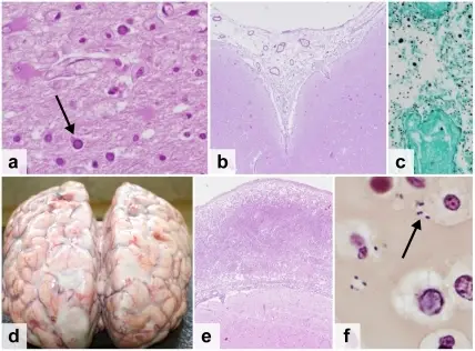
 Meningitis. Histopathology of bacterial meningitis: autopsy case of a person with pneumococcal meningitis showing inflammatory infiltrates of the pia mater consisting of neutrophil granulocytes (inset, higher magnification). Marvin 101. Not altered.CC BY-SA 3.0