Pleura pathology involves abnormalities of the pleura.
Pleura pathology includes:
- Pneumothorax
- Mesothelioma
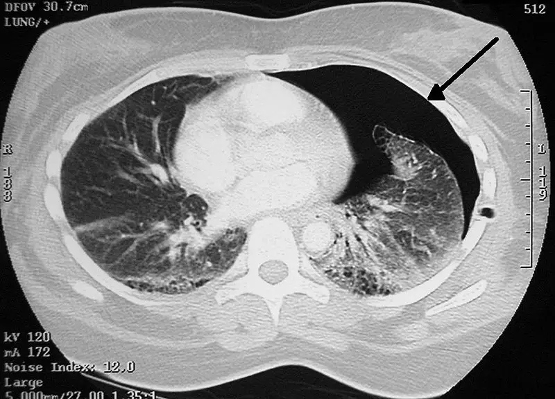
Pneumothorax
Pneumothorax is air buildup in the pleural space.
Spontaneous pneumothorax is because of rupture of an emphysematous bleb.
Pneumothorax leads to collapse of a portion of the lung.
The trachea shifts to the same side of the collapse.
When there is a penetrating chest wound, tension pneumothorax develops.
The pleural gap allows air in but not airflow.
The trachea is pulled in the opposite direction of the wound.
Pneumothorax is a medical emergency.
Pneumothorax is treated by inserting a chest tube.
 Pneumothorax. A chest tube placed on the right for a pneumothorax James Heilman, MD -Not altered CC BY-SA 4.0


Mesothelioma
Mesothelioma is a malignant neoplasm of mesothelial cells.
Mesothelioma is strongly linked to occupational exposure to asbestos.
Mesothelioma presents with:
- Chest pain
- Dyspnea
- Recurrent pleural effusions
 Mesothelioma. Coronal reformat of a CT of the chest in a patient with left sided mesothelioma. Note the extensive pleural mass with contraction of the left hemithorax. Frank Gaillard. CC BY-SA 3.0.