Restrictive Lung Disease Pathology Video
Restrictive Lung Diseases
Restrictive lung diseases are characterized by restricted filling of the lung where the FVC is more decreased than FEV1 resulting in a FEV1/FVC ratio of above 70%.
Restrictive lung diseases are most commonly caused by interstitial diseases of the lung and chest wall abnormalities like obesity.
 Different Examples of Cryobiopsy Showing UIP pattern.A) A low-magnification image showing dense scarring obliterating the alveolar architecture and abruptly alternating with relatively normal lung (patchy fibrosis). Some fibroblastic foci are visible even at this magnification for their pale-gray color. B) Fibroblastic focus better visualized at higher magnification. C) An area of honeycombing. Transbronchial lung cryobiopsy in the diagnosis of fibrotic interstitial lung diseases. Casoni GL, Tomassetti S, Cavazza A, Colby TV, Dubini A, Ryu JH, Carretta E, Tantalocco P, Piciucchi S, Ravaglia C, Gurioli C, Romagnoli M, Gurioli C, Chilosi M, Poletti V - PloS one (2014). Not Altered. CC.


Idiopathic Pulmonary Fibrosis (IPF)
Idiopathic pulmonary fibrosis (IPF) is characterized by the scarring or fibrosis of lung interstitium.
Although the etiology is unknown, idiopathic pulmonary fibrosis (IPF) is likely related to cyclical lung injury as injured pneumocytes induce fibrosis.
Causes of idiopathic pulmonary fibrosis (IPF) include:
- Cyclical lung injury
- Bleomycin
- Amiodarone
- Radiation therapy
Clinical features of idiopathic pulmonary fibrosis (IPF) include:
- Cough
- Progressive dyspnea and cough
Fibrosis on lung CT shows subpleural patches, which progress to diffuse fibrosis and eventually forms a ‘honeycomb’ pattern which is an indicator of end-stage pulmonary fibrosis.
The recommended treatment of idiopathic pulmonary fibrosis involves lung transplantation.
 Idiopathic Pulmonary Fibrosis. Not altered. CC


Pneumoconiosis
Pneumoconiosis is the form of interstitial fibrosis caused by occupational exposure to fibrogenic particles like asbestos, beryllium, and silica dusts.
The alveolar macrophages in the lungs engulf these particles inducing fibrosis.
 Pnemoconioses. High magnification micrograph of asbestosis of the lung, with characteristic ferruginous bodies and interstitial fibrosis. Lung biopsy. H&E stain. See also Image:Ferruginous_body.jpg - close-up of ferruginous bodies. Nephron. Not altered. CC BY-SA 3.0
Sarcoidosis
Sarcoidosis is a systemic disease characterized by growing granulomas in various body organs and is more prevalent in African American women.
Although its etiology is unknown sarcoidosis likely rises from the response of CD4+ helper T-cells to an unknown antigen.
The granulomas mostly affect the hilar lymph nodes and lungs, causing restrictive lung disease.
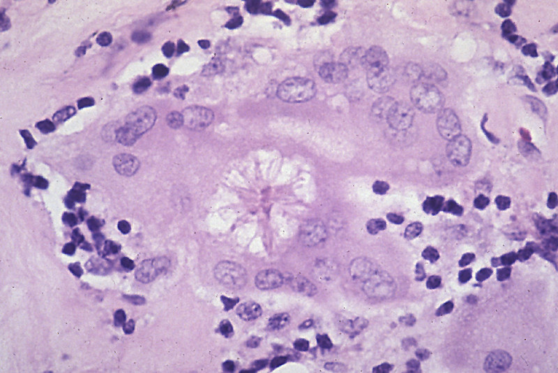
Histology of sarcoidosis includes naked granulomas and stellate inclusions (‘asteroid bodies’) are often seen within giant cells of the granulomas.
Other commonly affected tissues in sarcoidosis include:
- Uvea (uveitis)
- Skin (cutaneous nodules or erythema nodosum)
- Salivary glands
- Lacrimal glands
Clinical features of sarcoidosis include:
- Dyspnea
- Cough
Laboratory findings of sarcoidosis include:
- Elevated serum ACE
- Hypercalcemia
Steroids are the most preferred treatment although the condition often resolves on its own.
 Sarcoidosis


Hypersensitivity Pneumonitis
Hypersensitivity pneumonitis is mainly characterized by granulomatous reaction to inhaled organic antigens like pigeon breeder’s lung.
Common signs and symptoms of hypersensitivity pneumonitis include:
- Fever
- Cough
- Dyspnea
Symptoms of hypersensitivity pneumonitis typically occur hours after exposure.
Symptoms of hypersensitivity pneumonitis usually resolve after removal of the antigen.
Chronic exposure to the predisposing factor causing the hypersensitivity pneumonitis may result in interstitial fibrosis.
 Hypersensitivity Pneumonitis. High magnification micrograph of hypersensitivity pneumonitis showing granulomatous inflammation. Trichrome stain. Nephron - Not altered. CC BY-SA 3.0