Skeletal muscle pathology is the study of diseases and disorders that effect skeletal muscle.
In this section we highlight non neoplastic entities that effect skeletal muscle, which include:
- Dermatomyositis
- Polymyositis
- Muscular dystrophy (Duchenne muscular dystrophy & Becker muscular dystrophy)
- Neuromuscular junction disorders (Myasthenia gravis & Lambert-Eaton syndrome)
For neoplastic lesions that may affect skeletal muscle please refer to Soft Tissue Tumors Pathology Study Guide.
Dermatomyositis
Dermatomyositis is an inflammatory disorder of the skin and skeletal muscle.
Dermatomyositis is characterized by muscle weakness and Gottron’s papules which is a pathognomonic distinctive skin rash.
The exact cause of dermatomyositis is unknown.
Some cases of dermatomyositis are associated with carcinoma, for example, gastric carcinoma.
Clinical features of dermatomyositis include:
- Bilateral proximal muscle weakness (distal involvement can develop late in disease)
- Rash of the upper eyelids (heliotrope rash)
- Grotton’s papules which are red papules on the knuckles, elbows, and knees
- Malar rash
Laboratory findings of dermatomyositis include:
- Increased creatinine kinase
- Positive antinuclear antibody (ANA)
- Positive anti-Jo-1 antibody
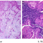
Histology of dermatomyositis will show perimysial inflammation (CD4+ T-cells) with perifascicular atrophy.
Corticosteroids are used for the treatment of dermatomyositis.
 Dermatomyositis. Gottron's papules on finger joints Elizabeth M. Dugan, Adam M. Huber, Frederick W. Miller, Lisa G. Rider - Not altered. CC BY-SA 3.0


Polymyositis
Polymyositis is an inflammatory disorder of skeletal muscle.
Polymyositis is a rare condition that weakens the muscles on both sides of the body.
Clinically, polymyositis resembles dermatomyositis but the skin is not involved.
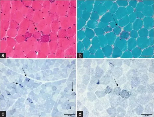
Histology of polymyositis will show endomysium inflammation (CD8+ T cells) with necrotic muscle fibers.
 Polymyositis. Inflammatory infiltrates in a muscle biopsy. Holes in the muscle cell vacuoles, deposits of abnormal proteins within the cells and in filamentous inclusions (hence the name inclusion body myositis} are clearly visible in the cellular interstitial space. Jensflorian -Not altered. CC BY-SA 3.0

Muscular Dystrophy
Muscular dystrophy is a degenerative disorder characterized by muscle wasting and replacement of skeletal muscle by adipose tissue.
The main types of muscular dystrophy to be aware of are:
- Duchenne muscular dystrophy (Duchenne is disastrous = worse prognosis)
- Becker muscular dystrophy (Becker is better = better prognosis)
Muscular dystrophy is caused by abnormal dystrophin.
The attachment of the muscle cytoskeleton to the extracellular matrix is made possible by dystrophin.
Mutations in dystrophin often occur spontaneously, and a high rate of mutation is predisposed by a large gene size.
Duchenne muscular dystrophy is due to deletion of dystrophin.
At around the age of 1-year-old, patients with Duchenne muscular dystrophy may have proximal muscular weakness that progresses to involve distal muscles.
Calf pseudohypertrophy is a characteristic finding of Duchenne muscular dystrophy.
Lab findings of Duchenne muscular dystrophy may show:
- Elevated lactic acid
- Elevated serum creatinine kinase is elevated
The heart is typically involved in Duchenne muscular dystrophy.
In Duchenne muscular dystrophy death typically results from cardiac or respiratory failure.
Becker muscular dystrophy is due to mutated dystrophin.
Becker muscular dystrophy clinically results in milder disease compared to Duchenne muscular dystrophy.
 Muscular Dystrophy. Muscular dystrophy is a genetic disorder where the muscle tissue wastes away and loses function. In the affected muscle (right), the tissue has become disorganized and the concentration of dystrophin (green), an important protein in normal muscle functioning, is greatly reduced. Cbenner - Not altered. CC BY-SA 3.0
Neuromuscular Junction Disorders
Myasthenia Gravis
Myasthenia gravis is a chronic autoimmune disorder due to autoantibodies against the postsynaptic acetylcholine receptor at the neuromuscular junction.
Myasthenia gravis is more commonly seen in women than men.
Clinical manifestations of Myasthenia gravis include:
- Muscle weakness that worsens with use
- Muscle weakness improves rest
- Ptosis (lid lag)
- Diplopia
Myasthenia gravis is associated with thymic hyperplasia and thymomas.
Thymectomy may improves symptoms.
Treatment of myasthenia gravis are anti-cholinesterase agents which help alleviate symptoms.
 Mysathenia Gravis. Strabismus in a person with myasthenia gravis trying to open their eyes. James Heilman, MD - Not altered. CC BY-SA 3.0


Lambert-Eaton Syndrome
Lambert-Eaton syndrome is due to antibodies directed against presynaptic calcium channels of the neuromuscular junction.
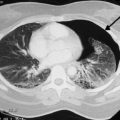
Lambert-Eaton syndrome may arise due to a paraneoplastic syndrome, most commonly due to small cell carcinoma of the lung.
Lambert-Eaton syndrome is associated with impaired acetylcholine release.
Firing of presynaptic calcium channels is required for acetylcholine release.
Clinical features of Lambert-Eaton syndrome include:
- Proximal muscle weakness that improves with use
Anticholinesterase agents do not cause any improvement of the symptoms due to Lambert-Eaton syndrome.
Lambert-Eaton syndrome may resolves with resection of the cancer if the cancer is the underlying cause.
 Lambert-Eaton Syndrome. Not altered. CC BY-SA 3.0